Regulatory Enzymes:- Allosteric Enzymes Undergo Conformational Changes in Response to Modulator Binding
As we saw in Chapter 5, allosteric proteins are those having “other shapes” or conformations induced by the binding of modulators. The same concept applies to certain regulatory enzymes, as conformational changes induced by one or more modulators interconvert more active and less-active forms of the enzyme. The modulators for allosteric enzymes may be inhibitory or stimulatory. Often the modulator is the substrate itself; regulatory enzymes for which substrate and modulator are identical are called homotropic. The effect is similar to that of O2 binding to hemoglobin (Chapter 5): binding of the ligand—or substrate, in the case of enzymes— causes conformational changes that affect the subsequent activity of other sites on the protein. When the modulator is a molecule other than the substrate, the enzyme is said to be heterotropic. Note that allosteric modulators should not be confused with uncompetitive and mixed inhibitors. Although the latter bind at a sec ond site on the enzyme, they do not necessarily mediate conformational changes between active and inactive forms, and the kinetic effects are distinct. The properties of allosteric enzymes are significantly different from those of simple nonregulatory enzymes. Some of the differences are structural. In addition to active sites, allosteric enzymes generally have one or more regulatory, or allosteric, sites for binding the modulator (Fig. 6–26). Just as an enzyme’s active site is specific for its substrate, each regulatory site is specific for its modulator. Enzymes with several modulators generally have different specific binding sites for each. In homotropic enzymes, the active site and regulatory site are the same. Allosteric enzymes are generally larger and more complex than nonallosteric enzymes. Most have two or more subunits. Aspartate transcarbamoylase, which catalyzes an early reaction in the biosynthesis of pyrimidine nucleotides (see Fig. 22–36), has 12 polypeptide chains organized into catalytic and regulatory subunits. Figure 6–27 shows the quaternary structure of this en zyme, deduced from x-ray analysis.
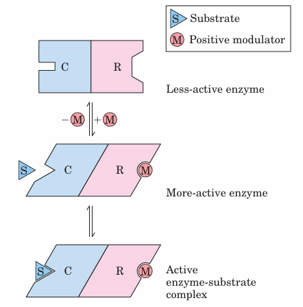
FIGURE 6–26 Subunit interactions in an allosteric enzyme, and in teractions with inhibitors and activators. In many allosteric enzymes the substrate binding site and the modulator binding site(s) are on different subunits, the catalytic (C) and regulatory (R) subunits, respectively. Binding of the positive (stimulatory) modulator (M) to its specific site on the regulatory subunit is communicated to the catalytic subunit through a conformational change. This change renders the catalytic subunit active and capable of binding the substrate (S) with higher affinity. On dissociation of the modulator from the regulatory subunit, the enzyme reverts to its inactive or less active form.
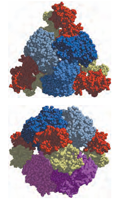
FIGURE 6–27 Two views of the regulatory enzyme aspartate transcarbamoylase. (Derived from PDB ID 2AT2.) This allosteric regulatory enzyme has two stacked catalytic clusters, each with three catalytic polypeptide chains (in shades of blue and purple), and three regula tory clusters, each with two regulatory polypeptide chains (in red and yellow). The regulatory clusters form the points of a triangle surround ing the catalytic subunits. Binding sites for allosteric modulators are on the regulatory subunits. Modulator binding produces large changes in enzyme conformation and activity. The role of this enzyme in nucleotide synthesis.








