Introduction and Bacterial Forms
 المؤلف:
Fritz H. Kayser
 المصدر:
Medical Microbiology -2005
 الجزء والصفحة:
 1-3-2016
 3355
3355
Introduction and Bacterial Forms
Bacterial cells are between 0.3 and 5 µm in size. They have three basic forms: cocci, straight rods, and curved or spiral rods. The nucleoid consists of a very thin, long, circular DNA molecular double strand that is not surrounded by a membrane. Among the nonessential genetic structures are the plasmids.
The cytoplasmic membrane harbors numerous proteins such as permeases, cell wall synthesis enzymes, sensor proteins, secretion system proteins, and, in aerobic bacteria, respiratory chain enzymes. The membrane is surrounded by the cell wall, the most important element of which is the supporting murein skeleton.
The cell wall of Gram-negative bacteria features a porous outer membrane into the outer surface of which the lipopolysaccharide responsible for the pathogenesis of Gram-negative infections is integrated. The cell wall of Gram-positive bacteria does not possess such an outer membrane. Its murein layer is thicker and contains teichoic acids and wall-associated proteins that contribute to the pathogenic process in Gram-positive infections.
Many bacteria have capsules made of polysaccharides that protect them from phagocytosis. Attachment pili or fimbriae facilitate adhesion to host cells. Motile bacteria possess flagella. Foreign body infections are caused by bacteria that form a biofilm on inert surfaces. Some bacteria produce spores, dormant forms that are highly resistant to chemical and physical noxae.
Bacteria differ from other single-cell microorganisms in both their cell structure and size, which varies from 0.3-5 im. Magnifications of 500- 1000x—close to the resolution limits of light microscopy—are required to obtain useful images of bacteria. Another problem is that the structures of objects the size of bacteria offer little visual contrast. Techniques like phase contrast and dark field microscopy, both of which allow for live cell observation, are used to overcome this difficulty. Chemical-staining techniques are also used, but the prepared specimens are dead.


- Simple staining. In this technique, a single staining substance, e.g., methylene blue, is used.
- Differential staining. Two stains with differing affinities to different bac¬teria are used in differential staining techniques, the most important of which is gram staining. Gram-positive bacteria stain blue-violet, Gram-negative bacteria stain red.
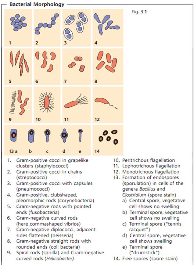
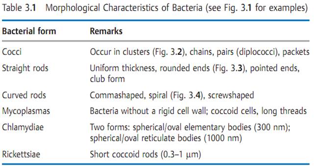
Three basic forms are observed in bacteria: spherical, straight rods, and curved rods (see Figs. 3.1).
 الاكثر قراءة في البكتيريا
 اخر الاخبار
اخبار العتبة العباسية المقدسة


