Platelet Plug Formation: Activation
 المؤلف:
Denise R. Ferrier
 المصدر:
Lippincott Illustrated Reviews: Biochemistry
 الجزء والصفحة:
 7-1-2022
 2086
2086
Platelet Plug Formation: Activation
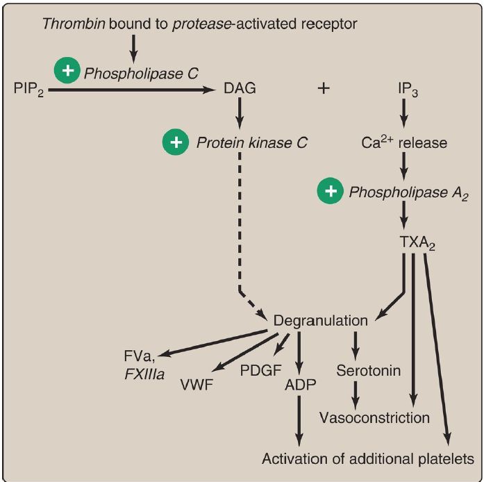
Once adhered to areas of injury, platelets get activated. Platelet activation involves morphologic (shape) changes and degranulation, the process by which platelets secrete the contents of their α and δ (or, dense) storage granules. Activated platelets also expose PS on their surface. The externalization of PS is mediated by a Ca2+-activated enzyme known as scramblase that disrupts the membrane asymmetry created by flippases . Thrombin is the most potent platelet activator. FIIa binds to and activates protease-activated receptors, a type of G protein–coupled receptor (GPCR), on the surface of platelets (Fig. 1). FIIa is primarily associated with Gq proteins, resulting in activation of phospholipase C and a rise in diacylglycerol (DAG) and inositol trisphosphate (IP3). [Note: Thrombomodulin, through its binding of FIIa, decreases the availability of FIIa for platelet activation .]

Figure 1: Platelet activation by thrombin. [Note: Protease-activated receptors are a type of G protein–coupled receptor.] PIP2 = phosphoinositol bisphosphate; DAG = diacylglycerol; IP3 = inositol trisphosphate; Ca2+ = calcium; TXA2 = thromboxane A2; ADP = adenosine diphosphate; PDGF = platelet-derived growth factor; VWF = von Willebrand factor; F = factor.
1. Degranulation: DAG activates protein kinase C, a key event for degranulation. IP3 causes the release of Ca2+ (from dense granules). The Ca2+ activates phospholipase A2, which cleaves membrane phospholipids to release arachidonic acid, the substrate for the synthesis of thromboxane A2 (TXA2) in activated platelets by cyclooxygenase-1
(COX-1) . TXA2 causes vasoconstriction, augments degranulation, and binds to platelet GPCR, causing activation of additional platelets. Recall that aspirin irreversibly inhibits COX and, consequently, TXA2 synthesis and is referred to as an antiplatelet drug.
Degranulation also results in release of serotonin and adenosine diphosphate (ADP) from dense granules. Serotonin causes vasoconstriction. ADP binds to GPCR on the surface of platelets, activating additional platelets. [Note: Some antiplatelet drugs, such as clopidogrel, are ADP-receptor antagonists.] Platelet-derived growth factor (involved in wound healing), VWF, FV, FXIII, and FI are among other proteins released from α granules. [Note: Platelet-activating factor (PAF), an ether phospholipid synthesized by a variety of cell types including endothelial cells and platelets, binds PAF receptors (GPCR) on the surface of platelets and activates them.]
2. Morphologic change: The change in shape of activated platelets from discoidal to spherical with pseudopod-like processes that facilitate platelet–platelet and platelet–surface interactions (Fig. 2) is initiated by the release of Ca2+ from dense granules. Ca2+ bound to calmodulin mediates the activation of myosin light chain kinase that phosphorylates the myosin light chain, resulting in a major reorganization of the platelet cytoskeleton.

Figure 2: Activated platelets undergo calcium (Ca2+)-initiated shape change.
 الاكثر قراءة في الكيمياء الحيوية
 اخر الاخبار
اخبار العتبة العباسية المقدسة


