


النبات

مواضيع عامة في علم النبات
الجذور - السيقان - الأوراق
النباتات الوعائية واللاوعائية
البذور (مغطاة البذور - عاريات البذور)
الطحالب
النباتات الطبية
الحيوان
مواضيع عامة في علم الحيوان
علم التشريح
التنوع الإحيائي
البايلوجيا الخلوية
الأحياء المجهرية
البكتيريا
الفطريات
الطفيليات
الفايروسات
علم الأمراض
الاورام
الامراض الوراثية
الامراض المناعية
الامراض المدارية
اضطرابات الدورة الدموية
مواضيع عامة في علم الامراض
الحشرات
التقانة الإحيائية
مواضيع عامة في التقانة الإحيائية
التقنية الحيوية المكروبية
التقنية الحيوية والميكروبات
الفعاليات الحيوية
وراثة الاحياء المجهرية
تصنيف الاحياء المجهرية
الاحياء المجهرية في الطبيعة
أيض الاجهاد
التقنية الحيوية والبيئة
التقنية الحيوية والطب
التقنية الحيوية والزراعة
التقنية الحيوية والصناعة
التقنية الحيوية والطاقة
البحار والطحالب الصغيرة
عزل البروتين
هندسة الجينات
التقنية الحياتية النانوية
مفاهيم التقنية الحيوية النانوية
التراكيب النانوية والمجاهر المستخدمة في رؤيتها
تصنيع وتخليق المواد النانوية
تطبيقات التقنية النانوية والحيوية النانوية
الرقائق والمتحسسات الحيوية
المصفوفات المجهرية وحاسوب الدنا
اللقاحات
البيئة والتلوث
علم الأجنة
اعضاء التكاثر وتشكل الاعراس
الاخصاب
التشطر
العصيبة وتشكل الجسيدات
تشكل اللواحق الجنينية
تكون المعيدة وظهور الطبقات الجنينية
مقدمة لعلم الاجنة
الأحياء الجزيئي
مواضيع عامة في الاحياء الجزيئي
علم وظائف الأعضاء
الغدد
مواضيع عامة في الغدد
الغدد الصم و هرموناتها
الجسم تحت السريري
الغدة النخامية
الغدة الكظرية
الغدة التناسلية
الغدة الدرقية والجار الدرقية
الغدة البنكرياسية
الغدة الصنوبرية
مواضيع عامة في علم وظائف الاعضاء
الخلية الحيوانية
الجهاز العصبي
أعضاء الحس
الجهاز العضلي
السوائل الجسمية
الجهاز الدوري والليمف
الجهاز التنفسي
الجهاز الهضمي
الجهاز البولي
المضادات الميكروبية
مواضيع عامة في المضادات الميكروبية
مضادات البكتيريا
مضادات الفطريات
مضادات الطفيليات
مضادات الفايروسات
علم الخلية
الوراثة
الأحياء العامة
المناعة
التحليلات المرضية
الكيمياء الحيوية
مواضيع متنوعة أخرى
الانزيمات

The pericardium
 المؤلف:
Harold Ellis,Vishy Mahadevan
المؤلف:
Harold Ellis,Vishy Mahadevan
 المصدر:
Clinical Anatomy Applied Anatomy for Students and Junior Doctors
المصدر:
Clinical Anatomy Applied Anatomy for Students and Junior Doctors
 الجزء والصفحة:
13th Edition , p30-32
الجزء والصفحة:
13th Edition , p30-32
 2025-02-25
 1213
1213


+
-
20
The heart and the roots of the great vessels are contained within the conical fibrous pericardium, the apex of which is fused with the adventitia of the great vessels and the base with the central tendon of the diaphragm. Anteriorly it is related to the body of the sternum, to which it is attached by the sternopericardial ligaments, the 3rd–6th costal cartilages and the anterior borders of the lungs. Posteriorly, it is related to the oesophagus, descending aorta and vertebra T5–T8, and on either side to the roots of the lungs, the mediastinal pleura and the phrenic nerves.
The pericardial cavity is the potential space between the visceral and parietal layers of the pericardium. Just like the pleural and peritoneal cavities, it is lubricated by a film of serous fluid. Following trauma it may fill with blood (haemopericardium).
The inner aspect of the fibrous pericardium is lined by the parietal layer of serous pericardium. This, in turn, is reflected around the roots of the great vessels to become continuous with the visceral layer or epicardium. The lines of pericardial reflexion are marked on the posterior surface of the heart (Fig. 1) by the oblique sinus, bounded by the inferior vena cava and the four pulmonary veins, which form a recess between the left atrium and the pericardium, and the transverse sinus between the superior vena cava and left atrium behind and the pulmonary trunk and aorta in front.
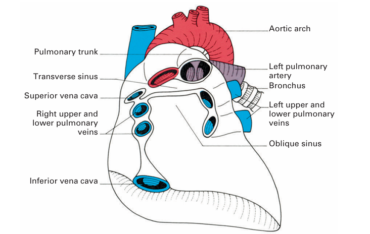
Fig1. The transverse and oblique sinuses of the pericardium. The heart has been removed from the pericardial sac, which is seen in anterior view.
 الاكثر قراءة في علم التشريح
 اخر الاخبار
اخبار العتبة العباسية المقدسة

الآخبار الصحية


 قسم الشؤون الفكرية يصدر كتاباً يوثق تاريخ السدانة في العتبة العباسية المقدسة
 "المهمة".. إصدار قصصي يوثّق القصص الفائزة في مسابقة فتوى الدفاع المقدسة للقصة القصيرة
 (نوافذ).. إصدار أدبي يوثق القصص الفائزة في مسابقة الإمام العسكري (عليه السلام)


