


النبات

مواضيع عامة في علم النبات
الجذور - السيقان - الأوراق
النباتات الوعائية واللاوعائية
البذور (مغطاة البذور - عاريات البذور)
الطحالب
النباتات الطبية
الحيوان
مواضيع عامة في علم الحيوان
علم التشريح
التنوع الإحيائي
البايلوجيا الخلوية
الأحياء المجهرية
البكتيريا
الفطريات
الطفيليات
الفايروسات
علم الأمراض
الاورام
الامراض الوراثية
الامراض المناعية
الامراض المدارية
اضطرابات الدورة الدموية
مواضيع عامة في علم الامراض
الحشرات
التقانة الإحيائية
مواضيع عامة في التقانة الإحيائية
التقنية الحيوية المكروبية
التقنية الحيوية والميكروبات
الفعاليات الحيوية
وراثة الاحياء المجهرية
تصنيف الاحياء المجهرية
الاحياء المجهرية في الطبيعة
أيض الاجهاد
التقنية الحيوية والبيئة
التقنية الحيوية والطب
التقنية الحيوية والزراعة
التقنية الحيوية والصناعة
التقنية الحيوية والطاقة
البحار والطحالب الصغيرة
عزل البروتين
هندسة الجينات
التقنية الحياتية النانوية
مفاهيم التقنية الحيوية النانوية
التراكيب النانوية والمجاهر المستخدمة في رؤيتها
تصنيع وتخليق المواد النانوية
تطبيقات التقنية النانوية والحيوية النانوية
الرقائق والمتحسسات الحيوية
المصفوفات المجهرية وحاسوب الدنا
اللقاحات
البيئة والتلوث
علم الأجنة
اعضاء التكاثر وتشكل الاعراس
الاخصاب
التشطر
العصيبة وتشكل الجسيدات
تشكل اللواحق الجنينية
تكون المعيدة وظهور الطبقات الجنينية
مقدمة لعلم الاجنة
الأحياء الجزيئي
مواضيع عامة في الاحياء الجزيئي
علم وظائف الأعضاء
الغدد
مواضيع عامة في الغدد
الغدد الصم و هرموناتها
الجسم تحت السريري
الغدة النخامية
الغدة الكظرية
الغدة التناسلية
الغدة الدرقية والجار الدرقية
الغدة البنكرياسية
الغدة الصنوبرية
مواضيع عامة في علم وظائف الاعضاء
الخلية الحيوانية
الجهاز العصبي
أعضاء الحس
الجهاز العضلي
السوائل الجسمية
الجهاز الدوري والليمف
الجهاز التنفسي
الجهاز الهضمي
الجهاز البولي
المضادات الميكروبية
مواضيع عامة في المضادات الميكروبية
مضادات البكتيريا
مضادات الفطريات
مضادات الطفيليات
مضادات الفايروسات
علم الخلية
الوراثة
الأحياء العامة
المناعة
التحليلات المرضية
الكيمياء الحيوية
مواضيع متنوعة أخرى
الانزيمات

Kinocilia-Uterine Tube
 المؤلف:
Kuehnel,W
المؤلف:
Kuehnel,W
 المصدر:
Color Atlas of Cytology, Histology, and Microscopic Anatomy
المصدر:
Color Atlas of Cytology, Histology, and Microscopic Anatomy
 الجزء والصفحة:
 31-7-2016
 2027
2027


+
-
20
Kinocilia-Uterine Tube
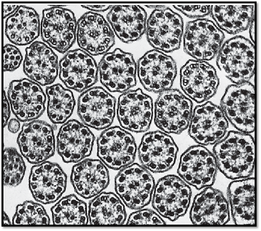
The detail structure of cilia is particularly impressive in cross-sections: there are two tubules in the center, and nine pairs of tubules (doublets) are located around them in the periphery (9 × 2 + 2 pattern). The peripheral twin tubules ( microtubule doublets, axoneme) are close to each other and share a common central wall. The wall consists of two or three protofilaments that are pre-dominantly compose d of the protein tubulin (α- and β-tubulin). This particular arrangement creates two different tubules within the twin tubules (doublet); only one of them, the A -microtubule , features a complete ring structure with 13 protofilaments. The B -tubule forms an incomplete ring. The B- tubule itself consists of only nine protofilaments. Three or four protofilaments in its structure belong to the A-ring. In contrast to the central microtubule pair, the A-tubules at the periphery have small “arms,” which reach to the adjacent pairs of microtubules. This figure shows these arms only vaguely. The arms consist of the protein dynein and ATPase. They play an important role in the motility of cilia. The two central microtubules are separate, side-by-side units. The peripheral axoneme are clearly connected to each other via the binding protein (nexin ). Axial view of the ciliate d cells from oviduct.
Electron microscopy; magnification: × 60 000
Longitudinal section through the kinocilia of a ciliate d epithelial cell from oviduct. Between the cilia are several sections through arcuate microvilli in different angles. Inside the cilia, microtubules form the longitudinal axis (axoneme), which consist of two central tubules (central tubule pair) and nine peripheral double tubules (doublets, each made of A - and B-tubules): 9 × 2 + 2 structure. The nine parallel outer axial tubules are connected to the central two tubules via spoke proteins. The two peripheral double tubules are different: in an axial section, tubule A forms a ring. In contrast, tubule B look s like an incomplete circle, like the letter C. The peripheral ciliary microtubules extend into the apical cytoplasm region. There, underneath the cell surface, they form basal bodies ( basal nodes, kinetosomes, cilia root ) . Kinetosomes form the cytoplasmic anchor places for microtubules. Their structure is that of centrioles. At the basal side, there is often a structure called rootstock , which shows a striation pattern. The rootstock contains the protein centrin . Kinetosomes are densely packed in richly ciliate d epithelium. This accounts for the layer of basal bodies ( basal nodule row ) that is visible in light microscopy.
1 Apical cytoplasm in a ciliated cell
Electron microscopy; magnification: × 30 000

References
Kuehnel, W.(2003). Color Atlas of Cytology, Histology, and Microscopic Anatomy. 4th edition . Institute of Anatomy Universitätzu Luebeck Luebeck, Germany . Thieme Stuttgar t · New York .
 الاكثر قراءة في علم الخلية
 اخر الاخبار
اخبار العتبة العباسية المقدسة

الآخبار الصحية


مواضيع ذات صلة
 قسم الشؤون الفكرية يصدر كتاباً يوثق تاريخ السدانة في العتبة العباسية المقدسة
 "المهمة".. إصدار قصصي يوثّق القصص الفائزة في مسابقة فتوى الدفاع المقدسة للقصة القصيرة
 (نوافذ).. إصدار أدبي يوثق القصص الفائزة في مسابقة الإمام العسكري (عليه السلام)


