
النبات

مواضيع عامة في علم النبات

الجذور - السيقان - الأوراق

النباتات الوعائية واللاوعائية

البذور (مغطاة البذور - عاريات البذور)

الطحالب

النباتات الطبية


الحيوان

مواضيع عامة في علم الحيوان

علم التشريح

التنوع الإحيائي

البايلوجيا الخلوية


الأحياء المجهرية

البكتيريا

الفطريات

الطفيليات

الفايروسات


علم الأمراض

الاورام

الامراض الوراثية

الامراض المناعية

الامراض المدارية

اضطرابات الدورة الدموية

مواضيع عامة في علم الامراض

الحشرات


التقانة الإحيائية

مواضيع عامة في التقانة الإحيائية


التقنية الحيوية المكروبية

التقنية الحيوية والميكروبات

الفعاليات الحيوية

وراثة الاحياء المجهرية

تصنيف الاحياء المجهرية

الاحياء المجهرية في الطبيعة

أيض الاجهاد

التقنية الحيوية والبيئة

التقنية الحيوية والطب

التقنية الحيوية والزراعة

التقنية الحيوية والصناعة

التقنية الحيوية والطاقة

البحار والطحالب الصغيرة

عزل البروتين

هندسة الجينات


التقنية الحياتية النانوية

مفاهيم التقنية الحيوية النانوية

التراكيب النانوية والمجاهر المستخدمة في رؤيتها

تصنيع وتخليق المواد النانوية

تطبيقات التقنية النانوية والحيوية النانوية

الرقائق والمتحسسات الحيوية

المصفوفات المجهرية وحاسوب الدنا

اللقاحات

البيئة والتلوث


علم الأجنة

اعضاء التكاثر وتشكل الاعراس

الاخصاب

التشطر

العصيبة وتشكل الجسيدات

تشكل اللواحق الجنينية

تكون المعيدة وظهور الطبقات الجنينية

مقدمة لعلم الاجنة


الأحياء الجزيئي

مواضيع عامة في الاحياء الجزيئي


علم وظائف الأعضاء


الغدد

مواضيع عامة في الغدد

الغدد الصم و هرموناتها

الجسم تحت السريري

الغدة النخامية

الغدة الكظرية

الغدة التناسلية

الغدة الدرقية والجار الدرقية

الغدة البنكرياسية

الغدة الصنوبرية

مواضيع عامة في علم وظائف الاعضاء

الخلية الحيوانية

الجهاز العصبي

أعضاء الحس

الجهاز العضلي

السوائل الجسمية

الجهاز الدوري والليمف

الجهاز التنفسي

الجهاز الهضمي

الجهاز البولي


المضادات الميكروبية

مواضيع عامة في المضادات الميكروبية

مضادات البكتيريا

مضادات الفطريات

مضادات الطفيليات

مضادات الفايروسات

علم الخلية

الوراثة

الأحياء العامة

المناعة

التحليلات المرضية

الكيمياء الحيوية

مواضيع متنوعة أخرى

الانزيمات
Granular (Rough) Endoplasmic Reticulum (rER)—Ergastoplasm
المؤلف:
Kuehnel,W
المصدر:
Color Atlas of Cytology, Histology, and Microscopic Anatomy
الجزء والصفحة:
3-8-2016
6519
Granular (Rough) Endoplasmic Reticulum (rER)—Ergastoplasm
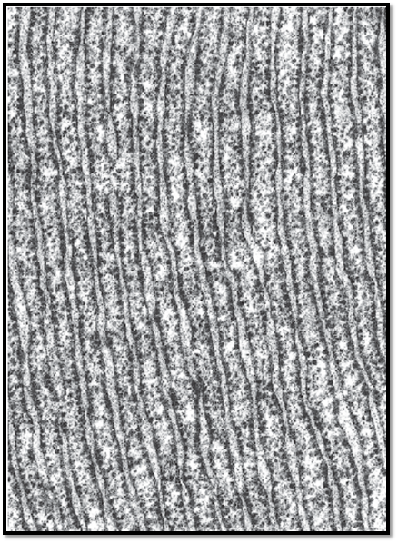
The endoplasmic reticulum (ER) is a continuous system of cell membranes, which are about 6 nm thick . Dependent on cell specialization and activity, the membranes occur in different forms, such as stacks or tubules. The ER double membranes may be smooth or have granules attached to their outer surfaces. These granules are about 25 nm in diameter and have been identified as membrane-bound ribosomes . Therefore, two types of ER exist: the granular or rough form ( rER, rough ER ) and the a granular or smooth form (sER, smooth ER ).
Paired multiplanar stacks of lamellae are one characteristic forms of rER. The membranes are narrowly spaced and spread over large parts of the cell. The two associated membranes in this matrix are 40–70 nm apart. When cells assume a storage function, these membranes move away from each other and thus form cisternae, with a lumen that may be several hundred nanometers wide.
Elaborate systems of rER membranes are found pre dominantly in cells that biosynthesize proteins . Proteins, which are synthesized on membranes of the rER, are mostly exported from the cell. They may be secreted from the cell (including hormones and digestive enzymes, etc.) or become part of intracellular vesicles ( membrane proteins ). The smooth endoplasmic reticulum eluded light microscopy.
The cisternae of the endoplasmic reticulum interconnect both with the perinuclear cisternae and the extracellular space. This picture shows ergastoplasm (rER) from an exocrine pancreas cell, which produces digestive enzymes.

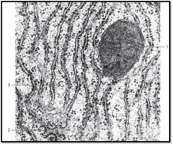
The granular endoplasmic reticulum (rER) exists not only in the form of strictly parallel-arrange d membrane stacks, which are shown in Figure 21 as transections. On the contrary, dependent on the specific function of a cell, rER is found in various forms and dimensions. The transition between granular and a granular ER can be continuous. In this figure, the rER presents as loosely packed stacks of cisternae with ribosomes attached to it like pearls on a string ( membrane-bound ribosomes). The figure shows a mitochondrion of crista-type 1 between the rER cisternae. There are also free ribosomes 3 and polysomes 2 in rosette configuration present in the cytoplasmic matrix. Such collocations of granular ER cisternae and adjacent free ribosomes are identical to the structures that are visible after staining with basophilic dyes in light microscopy preparations. Section from a rat hepatocyte.
1 Mitochondria with osmiophilic granula mitochondrialia
2 Polyribosomes
3 Free ribosomes

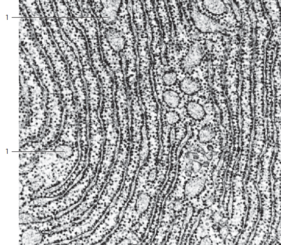
This figure shows rER double membranes, which are aligned in parallel and densely populated with ribosomes (ergastoplasm ). In the bottom part of the picture, the transections are slightly curve d. In some places, especially at their ends, the cisternae are distended into shapes resembling vacuoles, balloons or flasks. The rER content—consisting of proteins, which have all been synthesized on membrane-bound ribosomes—is frequently extracted during preparation, and therefore the rER cisternae appear empty.
However, in this section from a rat exocrine pancreatic cell, the narrow crevices as well as the large, distended cisternae show a fine granular or flocculent material. This material is the protein component of the definitive pancreatic excretion. The ergastoplasm—defined by the basophilic substance seen in light microscopy—is not solely due to membrane-bound ribosomes. The free ribosomes and polysomes in the cytoplasm also contribute to basophilia .

References
Kuehnel, W.(2003). Color Atlas of Cytology, Histology, and Microscopic Anatomy. 4th edition . Institute of Anatomy Universitätzu Luebeck Luebeck, Germany . Thieme Stuttgart · New York .
 الاكثر قراءة في علم الخلية
 اخر الاخبار
اخبار العتبة العباسية المقدسة

الآخبار الصحية


 قسم الشؤون الفكرية يصدر كتاباً يوثق تاريخ السدانة في العتبة العباسية المقدسة
 "المهمة".. إصدار قصصي يوثّق القصص الفائزة في مسابقة فتوى الدفاع المقدسة للقصة القصيرة
 (نوافذ).. إصدار أدبي يوثق القصص الفائزة في مسابقة الإمام العسكري (عليه السلام)


