
النبات

مواضيع عامة في علم النبات

الجذور - السيقان - الأوراق

النباتات الوعائية واللاوعائية

البذور (مغطاة البذور - عاريات البذور)

الطحالب

النباتات الطبية


الحيوان

مواضيع عامة في علم الحيوان

علم التشريح

التنوع الإحيائي

البايلوجيا الخلوية


الأحياء المجهرية

البكتيريا

الفطريات

الطفيليات

الفايروسات


علم الأمراض

الاورام

الامراض الوراثية

الامراض المناعية

الامراض المدارية

اضطرابات الدورة الدموية

مواضيع عامة في علم الامراض

الحشرات


التقانة الإحيائية

مواضيع عامة في التقانة الإحيائية


التقنية الحيوية المكروبية

التقنية الحيوية والميكروبات

الفعاليات الحيوية

وراثة الاحياء المجهرية

تصنيف الاحياء المجهرية

الاحياء المجهرية في الطبيعة

أيض الاجهاد

التقنية الحيوية والبيئة

التقنية الحيوية والطب

التقنية الحيوية والزراعة

التقنية الحيوية والصناعة

التقنية الحيوية والطاقة

البحار والطحالب الصغيرة

عزل البروتين

هندسة الجينات


التقنية الحياتية النانوية

مفاهيم التقنية الحيوية النانوية

التراكيب النانوية والمجاهر المستخدمة في رؤيتها

تصنيع وتخليق المواد النانوية

تطبيقات التقنية النانوية والحيوية النانوية

الرقائق والمتحسسات الحيوية

المصفوفات المجهرية وحاسوب الدنا

اللقاحات

البيئة والتلوث


علم الأجنة

اعضاء التكاثر وتشكل الاعراس

الاخصاب

التشطر

العصيبة وتشكل الجسيدات

تشكل اللواحق الجنينية

تكون المعيدة وظهور الطبقات الجنينية

مقدمة لعلم الاجنة


الأحياء الجزيئي

مواضيع عامة في الاحياء الجزيئي


علم وظائف الأعضاء


الغدد

مواضيع عامة في الغدد

الغدد الصم و هرموناتها

الجسم تحت السريري

الغدة النخامية

الغدة الكظرية

الغدة التناسلية

الغدة الدرقية والجار الدرقية

الغدة البنكرياسية

الغدة الصنوبرية

مواضيع عامة في علم وظائف الاعضاء

الخلية الحيوانية

الجهاز العصبي

أعضاء الحس

الجهاز العضلي

السوائل الجسمية

الجهاز الدوري والليمف

الجهاز التنفسي

الجهاز الهضمي

الجهاز البولي


المضادات الميكروبية

مواضيع عامة في المضادات الميكروبية

مضادات البكتيريا

مضادات الفطريات

مضادات الطفيليات

مضادات الفايروسات

علم الخلية

الوراثة

الأحياء العامة

المناعة

التحليلات المرضية

الكيمياء الحيوية

مواضيع متنوعة أخرى

الانزيمات
Chondral Osteogenesis-Finger
المؤلف:
Kuehnel, W
المصدر:
Color Atlas of Cytology, Histology, and Microscopic Anatomy
الجزء والصفحة:
5-1-2017
1782
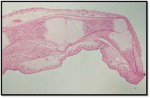
Chondral Osteogenesis-Finger
Longitudinal section through the index finger of a 6-month-old human embryo, with the base, middle and end of the limb to illustrate chondral osteogenesis (chondral, endochondral ossification), which begins in the dia-physis . Note the hyaline cartilage epiphyses. Chondral ossification is also referred to as indirect osteogenesis.
Stain: hematoxylin-eosin; magnification: × 18

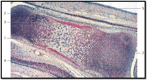
Chondral Osteogenesis-Finger
In contrast to the membranous, direct osteogenesis (ossification), the chondral osteogenesis requires an existing scaffold of hyaline cartilage lamellae ( cartilage model of ossification). While the cartilage is degrade d, it is replaced by bone tissue (substitute bone; indirect osteogenesis). Dependent on the lo-cation of their synthesis, there are perichondral and endochondral bones. The figure shows a longitudinal section through the middle phalanx of a finger. It presents the following details: the diaphysis 1 is in the center. Both the distal (left) and the proximal (right) epiphyses 2 still consist of hyaline cartilage (stained blue-violet). In the light zone between the two epiphyses, the diaphysis 1 , endochondral ossification has already started. The hyaline cartilage cells have changed into large spherical cells, and at the same time, calcium salts are deposited in the intercellular space. These areas can be recognized as small, branched, gray-blue lamellae. Osteoblasts reside in the connective tissue layer called perichondrium. They have built by membranous osteogenesis a sleeve of perichondral bone in the area of the dia-physis (here stained bright re d) 3 . The perichondrium is a form of connective tissue. The perichondral bone is a protective encasement for the bony structures in the area of the diaphysis. The two epiphyses are not yet affected by ossification.
1 Diaphysis
2 Epiphysis
3 Perichondral bone
4 Forming muscle cells
Stain: hematoxylin-eosin; magnification: × 15

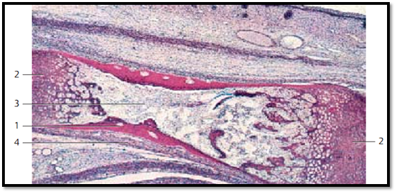
Chondral Osteogenesis—Finger
The perichondral bony sleeve 1 has become thicker by appositional growth . At the same time, it has expanded in the direction of the two epiphyses 2 . The cartilage cells in the diaphysis 3 have died, and the cartilage has disintegrated. In its place there is now vascular connective tissue, which has in-vaded from the outside. The tissue is mesenchymal in nature and is called primary bone marrow. Concurrently, small bony lamellae are created by chondral osteogenesis. In the direction of the epiphysis are rows of voluminous cartilage cells (column cartilage ), which stand out against the dormant cartilage tissue at the end of the joint .
1 Perichondral bone 3 Diaphysis
2 Epiphyseal cartilage 4 Periosteum
Stain: hematoxylin-eosin; magnification: × 12

References
Kuehnel, W.(2003). Color Atlas of Cytology, Histology, and Microscopic Anatomy. 4th edition . Institute of Anatomy Universitätzu Luebeck Luebeck, Germany . Thieme Stuttgart · New York .
 الاكثر قراءة في علم الخلية
 اخر الاخبار
اخبار العتبة العباسية المقدسة

الآخبار الصحية


 قسم الشؤون الفكرية يصدر كتاباً يوثق تاريخ السدانة في العتبة العباسية المقدسة
 "المهمة".. إصدار قصصي يوثّق القصص الفائزة في مسابقة فتوى الدفاع المقدسة للقصة القصيرة
 (نوافذ).. إصدار أدبي يوثق القصص الفائزة في مسابقة الإمام العسكري (عليه السلام)


