
النبات

مواضيع عامة في علم النبات

الجذور - السيقان - الأوراق

النباتات الوعائية واللاوعائية

البذور (مغطاة البذور - عاريات البذور)

الطحالب

النباتات الطبية


الحيوان

مواضيع عامة في علم الحيوان

علم التشريح

التنوع الإحيائي

البايلوجيا الخلوية


الأحياء المجهرية

البكتيريا

الفطريات

الطفيليات

الفايروسات


علم الأمراض

الاورام

الامراض الوراثية

الامراض المناعية

الامراض المدارية

اضطرابات الدورة الدموية

مواضيع عامة في علم الامراض

الحشرات


التقانة الإحيائية

مواضيع عامة في التقانة الإحيائية


التقنية الحيوية المكروبية

التقنية الحيوية والميكروبات

الفعاليات الحيوية

وراثة الاحياء المجهرية

تصنيف الاحياء المجهرية

الاحياء المجهرية في الطبيعة

أيض الاجهاد

التقنية الحيوية والبيئة

التقنية الحيوية والطب

التقنية الحيوية والزراعة

التقنية الحيوية والصناعة

التقنية الحيوية والطاقة

البحار والطحالب الصغيرة

عزل البروتين

هندسة الجينات


التقنية الحياتية النانوية

مفاهيم التقنية الحيوية النانوية

التراكيب النانوية والمجاهر المستخدمة في رؤيتها

تصنيع وتخليق المواد النانوية

تطبيقات التقنية النانوية والحيوية النانوية

الرقائق والمتحسسات الحيوية

المصفوفات المجهرية وحاسوب الدنا

اللقاحات

البيئة والتلوث


علم الأجنة

اعضاء التكاثر وتشكل الاعراس

الاخصاب

التشطر

العصيبة وتشكل الجسيدات

تشكل اللواحق الجنينية

تكون المعيدة وظهور الطبقات الجنينية

مقدمة لعلم الاجنة


الأحياء الجزيئي

مواضيع عامة في الاحياء الجزيئي


علم وظائف الأعضاء


الغدد

مواضيع عامة في الغدد

الغدد الصم و هرموناتها

الجسم تحت السريري

الغدة النخامية

الغدة الكظرية

الغدة التناسلية

الغدة الدرقية والجار الدرقية

الغدة البنكرياسية

الغدة الصنوبرية

مواضيع عامة في علم وظائف الاعضاء

الخلية الحيوانية

الجهاز العصبي

أعضاء الحس

الجهاز العضلي

السوائل الجسمية

الجهاز الدوري والليمف

الجهاز التنفسي

الجهاز الهضمي

الجهاز البولي


المضادات الميكروبية

مواضيع عامة في المضادات الميكروبية

مضادات البكتيريا

مضادات الفطريات

مضادات الطفيليات

مضادات الفايروسات

علم الخلية

الوراثة

الأحياء العامة

المناعة

التحليلات المرضية

الكيمياء الحيوية

مواضيع متنوعة أخرى

الانزيمات
Biotechnological Applications of Magnetic Nanoparticles
المؤلف:
John M Walker and Ralph Rapley
المصدر:
Molecular Biology and Biotechnology 5th Edition
الجزء والصفحة:
2-12-2020
1168
Biotechnological Applications of Magnetic Nanoparticles
Magnetic particles have shown considerable promise as probes for magnetic resonance imaging, as they provide strong contrast effects to surrounding tissues. These effects can be understood in terms of their influence on the spin–spin relaxation times (T2) of surrounding water molecules. Conventional iron oxide contrast agents such as the superparamagnetic iron oxides or the cross-linked iron oxides are of limited utility due to their poor magnetic contrast effects. New nanoparticles systems such as MEIO nanoparticles have high and tunable mass magnetization values capable of enhancing T2 relaxation times. The importance of nanoscaling laws in designing optimal MEIO systems can be seen in the size dependence of the relaxivity coefficient (r2), a direct indication of contrast enhancement. In 4nm MEIO particles, the relaxivity coefficient is 78mM-1 s-1, but increases to 106, 130 and 218mM-1 s-1 for 6, 9 and 12nm nanoparticles, respectively. Using dopants, the efficacy of these particles can also be tuned by changes in their composition. A 12nm Mn-doped MEIO with the
highest magnetization values of 110 emu g-1 (Mn+Fe) exhibits the best MR contrast with an r2 of 358mM-1 s-1. Other metal-doped MEIOs have values of 101 emu g-1 (Fe) for all Fe, 99 emu g-1 (Co+Fe) for Codoped and 85 emu g-1 (Ni+Fe) for Ni-substituted with r2 values of 218, 172 and 152mM-1 s-1, respectively.
Given that the r2 of the Mn–MEIO particle is six times higher that of most conventional molecular MR contrast imaging agents, these properties have been translated into effective enhancements for the ultrasensitive detection of in vivo biological targets. Using Mn–MEIO particles conjugated with Herceptin and injected into the tail vein of a
mouse, a small HER2/nue cancer was selectively detected by MRI imaging. In contrast, the same tumor was undetectable using convention cross-linked iron oxide–Herceptin conjugate.
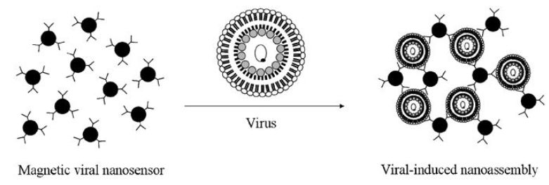
Although magnetic beads have long been widely used for magneticbased sensing and separations, problems still exist with these methods due to low magnetic susceptibility and considerable magnetic inhomogeneity. Magnetic nanoparticles can offer a solution to these problems. Superparamagnetic iron oxide nanoparticles have been used in a diagnostic format for the detection of virus particles. The principle of this assay is that the target virus acts to cross-link SPIO nanoparticles derivatized with targeting antibodies, forming larger assemblies with increased relaxation enhancements (Figure 1).

Figure 1. Antibody-functionalized nanoparticles induce the formation of nanoassemblies in the presence of a virus. This allows for viral detection by measuring the change in spin–spin relaxation times of surrounding molecules brought about nanoassembly formation.
In proof-of-concept experiments, the formation of the virus aggregated nanoassemblies was confirmed by light scattering experiments. When the samples were examined by MRI, the virus particles were detected in concentrations as low as five particles in 10 ml of biological sample. It is likely that detection limits could be further enhanced with a shift to MEIO particles and/or greater magnetic field strength.
Magnetic nanoparticles can also enhance magnetophoretic separations.
When a magnetic field is applied perpendicular to the flow direction of a microfluidic channel, magnetic particles experience a magnetic force which drives their lateral movement to a given velocity. The velocity of that magnetophoretic lateral movement (vlat) is proportional to the magnetic susceptibility of the particle and the square of the particle radius. The magnetic susceptibility of the nanoparticles is a key component of velocity control and consequently the achievement of good magnetophoretic separations.
Allergy tests require the quantitative detection of allergen-specific antibodies (IgE) in the serum of a patient suffering from allergies. Unfortunately, the IgE concentrations in allergy patients are usually low, driving the need for highly sensitive detection methods. To demonstrate the efficacy of magnetophoretic separation and sensing of dust mite IgE antibodies, microbeads coated with mite allergen from Dermotophagiodes farina were first mixed with target IgE (Figure 2).
To this solution, secondary anti-human IgE-coated MEIO nanoparticles were added. The resulting solution was injected into the microchannel of a magnetophoretic separator. At high concentrations of the target IgE, significant lateral movement of the beads was achieved (vlat=15 μms-1).
At lower concentrations of target, reduced (vlat=2 μms-1) or negligible lateral movement was detected. This is consistent with the idea that in this nanohybrid sandwich assay, higher concentrations of specific IgE result in more MEIO nanoparticles binding on to the microbeads. In sera, quantitative detection of target IgEs was achieved at the subpicomolar levels (~500 fM) when using a target IgE concentration versus lateral velocity calibration curve.

Figure 2. Sandwich assay demonstrating the detection and separation of specific allergen antibodies using antibody-functionalized magnetic nanoparticles. Reprinted with permission from Jun et al., J. Am. Chem. Soc., 2008, 41, 179. Copyright 2008 American Chemical Society.
Researchers are beginning to achieve a better understanding of the nanoscaling laws for magnetism. From these studies, it is apparent that size, shape and composition have a tremendous impact on magnetic parameters such as coercivity and magnetization values. Using these tunable properties, magnetic particles have begun to find their way into a variety of biotechnology applications. Research and discovery will lead to continued improvements in MRI, biosensing and magnetic separations and new advances in next-generation drug delivery and hyperthermia treatments.
 الاكثر قراءة في مفاهيم التقنية الحيوية النانوية
 اخر الاخبار
اخبار العتبة العباسية المقدسة

الآخبار الصحية


 قسم الشؤون الفكرية يصدر كتاباً يوثق تاريخ السدانة في العتبة العباسية المقدسة
 "المهمة".. إصدار قصصي يوثّق القصص الفائزة في مسابقة فتوى الدفاع المقدسة للقصة القصيرة
 (نوافذ).. إصدار أدبي يوثق القصص الفائزة في مسابقة الإمام العسكري (عليه السلام)


