

النبات

مواضيع عامة في علم النبات

الجذور - السيقان - الأوراق

النباتات الوعائية واللاوعائية

البذور (مغطاة البذور - عاريات البذور)

الطحالب

النباتات الطبية


الحيوان

مواضيع عامة في علم الحيوان

علم التشريح

التنوع الإحيائي

البايلوجيا الخلوية


الأحياء المجهرية

البكتيريا

الفطريات

الطفيليات

الفايروسات


علم الأمراض

الاورام

الامراض الوراثية

الامراض المناعية

الامراض المدارية

اضطرابات الدورة الدموية

مواضيع عامة في علم الامراض

الحشرات


التقانة الإحيائية

مواضيع عامة في التقانة الإحيائية


التقنية الحيوية المكروبية

التقنية الحيوية والميكروبات

الفعاليات الحيوية

وراثة الاحياء المجهرية

تصنيف الاحياء المجهرية

الاحياء المجهرية في الطبيعة

أيض الاجهاد

التقنية الحيوية والبيئة

التقنية الحيوية والطب

التقنية الحيوية والزراعة

التقنية الحيوية والصناعة

التقنية الحيوية والطاقة

البحار والطحالب الصغيرة

عزل البروتين

هندسة الجينات


التقنية الحياتية النانوية

مفاهيم التقنية الحيوية النانوية

التراكيب النانوية والمجاهر المستخدمة في رؤيتها

تصنيع وتخليق المواد النانوية

تطبيقات التقنية النانوية والحيوية النانوية

الرقائق والمتحسسات الحيوية

المصفوفات المجهرية وحاسوب الدنا

اللقاحات

البيئة والتلوث


علم الأجنة

اعضاء التكاثر وتشكل الاعراس

الاخصاب

التشطر

العصيبة وتشكل الجسيدات

تشكل اللواحق الجنينية

تكون المعيدة وظهور الطبقات الجنينية

مقدمة لعلم الاجنة


الأحياء الجزيئي

مواضيع عامة في الاحياء الجزيئي


علم وظائف الأعضاء


الغدد

مواضيع عامة في الغدد

الغدد الصم و هرموناتها

الجسم تحت السريري

الغدة النخامية

الغدة الكظرية

الغدة التناسلية

الغدة الدرقية والجار الدرقية

الغدة البنكرياسية

الغدة الصنوبرية

مواضيع عامة في علم وظائف الاعضاء

الخلية الحيوانية

الجهاز العصبي

أعضاء الحس

الجهاز العضلي

السوائل الجسمية

الجهاز الدوري والليمف

الجهاز التنفسي

الجهاز الهضمي

الجهاز البولي


المضادات الميكروبية

مواضيع عامة في المضادات الميكروبية

مضادات البكتيريا

مضادات الفطريات

مضادات الطفيليات

مضادات الفايروسات

علم الخلية

الوراثة

الأحياء العامة

المناعة

التحليلات المرضية

الكيمياء الحيوية

مواضيع متنوعة أخرى

الانزيمات
Phylum: Zygomycota
المؤلف:
Kayser, F. H
المصدر:
Medical Microbiology
الجزء والصفحة:
17-11-2015
5166
Phylum: Zygomycota
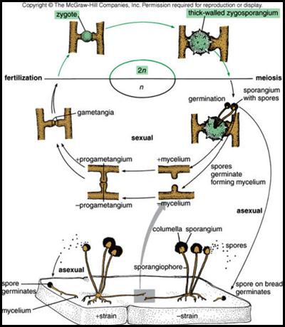
This phylum contains about 600 described species that are mostly terrestrial in habitat, living in soil or on decaying plant or animal material. This division has coenocytic mycelium, asexual reproduction is by non- motile spores (=sporangiospores) that are produced in sporangia borne on stalks (=sporangiophores). Zygomycota, like all true fungi, produce cell walls containing chitin. They grow primarily as mycelia, or filaments of long cells called hyphae. Their name is derived from their way of reproducing sexually. The principal characteristic that distinguishes this class is the production of a thick walled resting spore called a zygospore. The zygospore develops inside a zygosporangium that is formed after fusion or conjugation - of morphologically similar two gametangia (structures containing gametes).
Class: Zygomycetes
Characteristics of the class is the same as those of the phylum.
Order Mucorales:
This is widespread group of moulds occurring as furry growths on damp bread, rotting fruit, and especially on dung in humid weather • Examples: Rhizopus, Phycomyces, Mucor and Pilobolus
Mucoraceae
Rhizopus stolonifer: (The black bread mould)
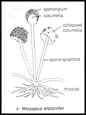
One of the commonest mucoraceous mould, it is abundant on damp bread and rotting fruit. the sporangia appear black. This mould produce fast- growing aerial hyphae (stolon) which arch over and touch down on a solid surface a head of the main mycelium, close to its point contact the stolon forms a tuft of root- like hyphae ( rhizoids).. From the region where the rhizoids formed, one to several short, stiff sporangiophores are formed. the sporangiospore released when the wall of the columellate sporangium disintegrates. Under favorable conditions a spore germinates by germ tube which develops into a fluffy, many branched, white, aerial mycelium. sexual reproduction is by mean of gametangial copulation. Colonies are fast growing and cover an agar surface with a dense cottony growth that is at first white becoming grey or yellowish brown with sporulation.
Mucor sp.
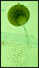
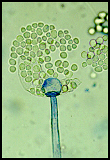
Colonies are very fast growing, cottony to fluffy, white to yellow, becoming dark grey with the development of sporangia. Sporangiophores are erect, simple or branched, forming large terminal, globose to spherical, multi-spored sporangia, with well developed subtending columellae. A conspicuous collarette (remnants of the sporangial wall) is usually visible at the base of the columella following sporangiospore dispersal. Sporangiospores are hyaline, grey or brownish. Stolons and rhizoids are absent, however, zygospores may be present.
 |
 |
Pilobolaceae:
Pilobolus :
Is a common inhabitant of horse and cow dung. It grows very rapidly, sporangiophores is unbranched, and have a unique explosive dispersal mechanism. Beneath the black apical sporangium is a lens-like subsporangial vesicle, with a light-sensitive `retina' at its base that controls the growth of the sporangiophore ,aiming it toward any light source. In a word, it is phototropic. Osmotically active compounds cause pressure in the sporangiophore and the subsporangial vesicle to build up until it is more than 100 pounds per square inch (7 kilograms per square centimetre). This eventually causes the vesicle to explode, hurling the black sporangium away to a distance of up to 2 metres, directly toward the light. The mucilaginous contents of the subsporangial vesicle go with the sporangium, and glue it to whatever it lands on
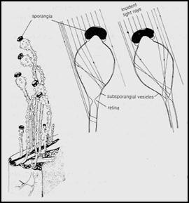
Live material:
Look at the available dung samples kept in large glass culture dishes. Use a dissection microscope. These will be kept in the fume hood with a heavy cover to prevent desiccation. Also a number of plates will be set up with water agar and small quantities of rabbit dung. It is expected that a variety of fungal types will develop on the dung and these plates should be examined at regular intervals (3 day) under a dissecting microscope.
 الاكثر قراءة في الفطريات
 اخر الاخبار
اخبار العتبة العباسية المقدسة

الآخبار الصحية


 قسم الشؤون الفكرية يصدر كتاباً يوثق تاريخ السدانة في العتبة العباسية المقدسة
 "المهمة".. إصدار قصصي يوثّق القصص الفائزة في مسابقة فتوى الدفاع المقدسة للقصة القصيرة
 (نوافذ).. إصدار أدبي يوثق القصص الفائزة في مسابقة الإمام العسكري (عليه السلام)


