تاريخ الفيزياء

علماء الفيزياء


الفيزياء الكلاسيكية

الميكانيك

الديناميكا الحرارية


الكهربائية والمغناطيسية

الكهربائية

المغناطيسية

الكهرومغناطيسية


علم البصريات

تاريخ علم البصريات

الضوء

مواضيع عامة في علم البصريات

الصوت


الفيزياء الحديثة


النظرية النسبية

النظرية النسبية الخاصة

النظرية النسبية العامة

مواضيع عامة في النظرية النسبية

ميكانيكا الكم

الفيزياء الذرية

الفيزياء الجزيئية


الفيزياء النووية

مواضيع عامة في الفيزياء النووية

النشاط الاشعاعي


فيزياء الحالة الصلبة

الموصلات

أشباه الموصلات

العوازل

مواضيع عامة في الفيزياء الصلبة

فيزياء الجوامد


الليزر

أنواع الليزر

بعض تطبيقات الليزر

مواضيع عامة في الليزر


علم الفلك

تاريخ وعلماء علم الفلك

الثقوب السوداء


المجموعة الشمسية

الشمس

كوكب عطارد

كوكب الزهرة

كوكب الأرض

كوكب المريخ

كوكب المشتري

كوكب زحل

كوكب أورانوس

كوكب نبتون

كوكب بلوتو

القمر

كواكب ومواضيع اخرى

مواضيع عامة في علم الفلك

النجوم

البلازما

الألكترونيات

خواص المادة


الطاقة البديلة

الطاقة الشمسية

مواضيع عامة في الطاقة البديلة

المد والجزر

فيزياء الجسيمات


الفيزياء والعلوم الأخرى

الفيزياء الكيميائية

الفيزياء الرياضية

الفيزياء الحيوية

الفيزياء العامة


مواضيع عامة في الفيزياء

تجارب فيزيائية

مصطلحات وتعاريف فيزيائية

وحدات القياس الفيزيائية

طرائف الفيزياء

مواضيع اخرى
X-RAYS
المؤلف:
S. Gibilisco
المصدر:
Physics Demystified
الجزء والصفحة:
484
5-11-2020
2041
X-RAYS
The x-ray spectrum consists of EM energy at wavelengths from approximately 1 nm down to 0.01 nm. (Various sources disagree somewhat on the dividing line between the hard-UV and x-ray regions.) Proportionately, the x-ray spectrum is large compared with the visible range.
X-rays were discovered accidentally in 1895 by a physicist named Wilhelm Roentgen during experiments involving electric currents in gases at low pressure. If the current was sufficiently intense, the high-speed electrons produced mysterious radiation when they struck the anode (positively charged electrode) in the tube. The rays were called x-rays because of their behavior, never before witnessed. The rays were able to penetrate barriers opaque to visible light and UV. A phosphor-coated object happened to be in the vicinity of the tube containing the gas, and Roentgen noticed that the phosphor glowed. Subsequent experiments showed that the rays possessed so much penetrating power that they passed through the skin and muscles in the human hand, casting shadows of the bones on a phosphor-coated surface. Photographic film could be exposed in the same way.
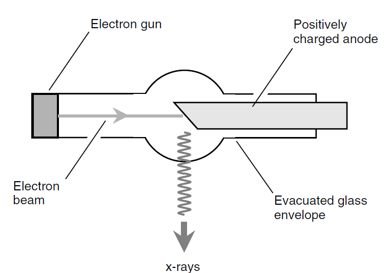
Modern x-ray tubes operate by accelerating electrons to high speed and forcing them to strike a heavy metal anode (usually made of tungsten). A simplified functional diagram of an x-ray tube of the sort used by dentists to locate cavities in your teeth is shown in Fig. 1.
As the wavelengths of x-rays become shorter and shorter, it becomes increasingly difficult to direct and focus them. This is so because of the penetrating power of the short-wavelength rays. A piece of paper with a tiny hole works very well for UV photography; in the x-ray spectrum, the radiation passes right through the paper. Even aluminum foil is relatively

Fig. 1. Functional diagram of an x-ray tube.
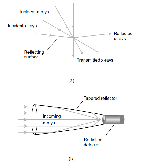
transparent to x-rays. However, if x-rays land on a reflecting surface at a nearly grazing angle, and if the reflecting surface is made of suitable material, some degree of focusing can be realized. The shorter the wavelength of the x-rays, the smaller the angle of incidence, measured relative to the surface (not the normal), must be if reflection is to take place. At the shortest x-ray wavelengths, the angle must be smaller than 1° of arc. This grazing reflection effect is shown in Fig. 2a. A rough illustration of how a high-resolution x-ray observing device achieves its focusing is shown in part b. The focusing mirror is tapered in the shape of an elongated paraboloid. As parallel x-rays enter the aperture of the reflector, they strike its inner surface at a grazing angle. The x-rays are brought to a focal point, where a radiation counter or detector can be placed.
X-rays cause ionization of living tissue. This effect is cumulative and can result in damage to cells over a period of years. This is why x-ray technicians in doctors’ and dentists’ offices work behind a barrier lined with lead. Otherwise, these personnel would be subjected to dangerous cumulative doses of x radiation. It only takes a few millimeters of lead to block virtually all x-rays. Less dense metals and other solids also can block x-rays, but these must be thicker. The important factor is the amount of mass through which the radiation must pass. Sheer physical displacement also can reduce the intensity of x radiation, which diminishes according to the square of the distance. However, it isn’t practical for most doctors or dentists to work in offices large enough to make this a viable alternative.

Fig. 2. (a) x-rays are reflected from a surface only when they strike at a grazing angle. (b) A functional diagram of an x-ray focusing and observing device.
 الاكثر قراءة في الكهرومغناطيسية
 اخر الاخبار
اخبار العتبة العباسية المقدسة

الآخبار الصحية


 قسم الشؤون الفكرية يصدر كتاباً يوثق تاريخ السدانة في العتبة العباسية المقدسة
 "المهمة".. إصدار قصصي يوثّق القصص الفائزة في مسابقة فتوى الدفاع المقدسة للقصة القصيرة
 (نوافذ).. إصدار أدبي يوثق القصص الفائزة في مسابقة الإمام العسكري (عليه السلام)


