آخر المواضيع المضافة

 النبات
الحيوان
الأحياء المجهرية
علم الأمراض
التقانة الإحيائية
التقنية الحيوية المكروبية
التقنية الحياتية النانوية
علم الأجنة
الأحياء الجزيئي
علم وظائف الأعضاء
الغدد
المضادات الحيوية
النبات
الحيوان
الأحياء المجهرية
علم الأمراض
التقانة الإحيائية
التقنية الحيوية المكروبية
التقنية الحياتية النانوية
علم الأجنة
الأحياء الجزيئي
علم وظائف الأعضاء
الغدد
المضادات الحيوية| Types of Collagen |


|
|
Read More
Date: 5-9-2021
Date: 7-12-2021
Date: 7-11-2021
|
Types of Collagen
The collagen superfamily of proteins includes >25 collagen types as well as additional proteins that have collagen-like domains. The three polypeptide α chains are held together by interchain hydrogen bonds. Variations in the amino acid sequence of the α chains result in structural components that are about the same size (~1,000 amino acids long) but with slightly different properties. These α chains are combined to form the various types of collagen found in the tissues. For example, the most common collagen, type I, contains two chains called α1 and one chain called α2 (α12α2), whereas type II collagen contains three α1 chains (α13). The collagens can be organized into three groups, based on their location and functions in the body (Fig. 1).
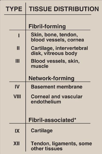
Figure 2 : The most abundant types of collagen. [Note: *Fibril-associated collagens with interrupted triple helices are known as FACIT.]
1. Fibril-forming collagens: Types I, II, and III are the fibrillar collagens and have the rope-like structure described above for a typical collagen molecule. In the electron microscope, these linear polymers of fibrils have characteristic banding patterns, reflecting the regular staggered packing of the individual collagen molecules in the fibril (Fig. 2). Type I collagen fibers (composed of collagen fibrils) are found in supporting elements of high tensile strength (for example, tendons and corneas), whereas fibers formed from type II collagen molecules are restricted to cartilaginous structures. The fibers derived from type III collagen are prevalent in more distensible tissues such as blood vessels.
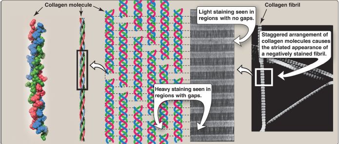
Figure 2: Collagen fibrils at right have a characteristic banding pattern, reflecting the regularly staggered packing of the individual collagen molecules in the fibril.
2. Network-forming collagens: Types IV and VIII form a three-dimensional mesh, rather than distinct fibrils (Fig. 3). For example, type IV molecules assemble into a sheet or meshwork that constitutes a major part of basement membranes.
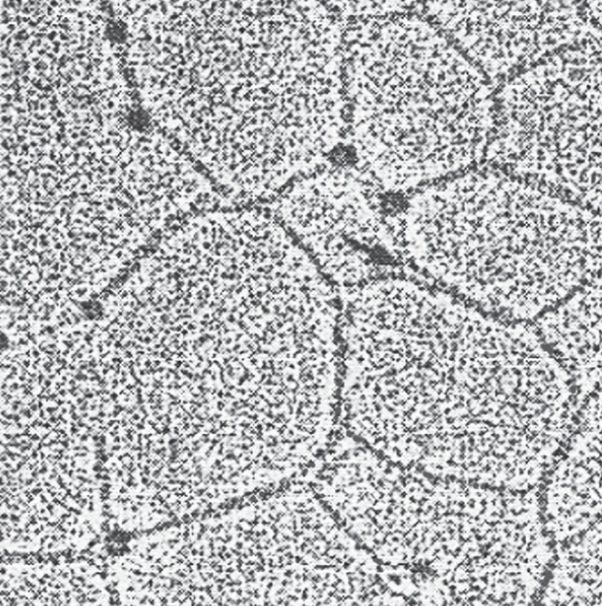
Figure 3: Electron micrograph of a polygonal network formed by association of collagen type IV monomers.
Basement membranes are thin, sheet-like structures that provide mechanical support for adjacent cells and function as a semipermeable filtration barrier to macromolecules in organs such as the kidney and the lung.
3. Fibril-associated collagens: Types IX and XII bind to the surface of collagen fibrils, linking these fibrils to one another and to other components in the ECM (see Fig. 1).


|
|
|
|
تحذير من "عادة" خلال تنظيف اللسان.. خطيرة على القلب
|
|
|
|
|
|
|
دراسة علمية تحذر من علاقات حب "اصطناعية" ؟!
|
|
|
|
|
|
|
العتبة العباسية المقدسة تحذّر من خطورة الحرب الثقافية والأخلاقية التي تستهدف المجتمع الإسلاميّ
|
|
|