


 النبات
 الحيوان
 الأحياء المجهرية
 علم الأمراض
 التقانة الإحيائية
 التقنية الحيوية المكروبية
 التقنية الحياتية النانوية
 علم الأجنة
 الأحياء الجزيئي
 علم وظائف الأعضاء
 الغدد
الغدد
 المضادات الحيوية
|
Read More
Date: 2025-01-16
Date: 2024-12-21
Date: 6-11-2015
|
A Concise History of Immunology
The role of smallpox in the development of vaccination
The concept of immunity from disease dates back at least to Greece in the 5th century BC. Thucydides wrote of individuals who recovered from the plague, which was raging in Athens at the time. These individuals, who had already contracted the disease, recovered and became “immune” or “exempt.” However, the earliest recognized attempt to intentionally induce immunity to an infectious disease was in the 10th century in China, where smallpox was endemic. The process of “variolation” involved exposing healthy people to material from the lesions caused by the disease, either by putting it under the skin, or, more often, inserting powdered scabs from smallpox pustules into the nose. Variolation was known and practiced frequently in the Ottoman Empire, where it had been introduced by Circassian traders around 1670. Unfortunately, because there was no standardization of the inoculum, the variolation occasionally resulted in death or disfigurement from smallpox, thus limiting its acceptance.
Variolation later became popular in England, mainly due to the efforts of Lady Mary Wortley Montague who survived smallpox but who lost a brother to it. Lady Montague was married to Lord Edward Wortley Montague, the ambassador to the Sublime Porte of the Ottomans in Istanbul. While in Istanbul, Lady Montague observed the practice of variolation. Determined not to have her family suffer as she had, she directed the surgeon of the Embassy to learn the technique and, in March 1718, to variolate her five year-old son. After her return to England, she promoted the technique, and had her surgeon variolate her four-year old daughter in the presence of the king’s physician. The surgeon, Charles Maitland, was given leave to perform what came to be known as the Royal Experiment, in which he variolated six condemned prisoners who later survived. By these and other experiments, the safety of the procedure was established, and two of the king’s grandchildren were variolated on April 17, 1722. After this, the practice of variolation spread rapidly throughout England in the 1740s and then to the American colonies.
Edward Jenner and the development of the first safe vaccine for smallpox
Although Jenner is rightly celebrated for his development of cowpox as a safe vaccine for smallpox, he was not the first to make use of a relatively non-pathogenic virus to induce immunity. In 1774, Benjamin Jesty, a farmer, inoculated his wife with the vaccinia virus obtained from “farmer Elford of Chittenhall, near Yetminster.” In 1796, Jenner inoculated James Phipps with material obtained from a cowpox lesion that appeared on the hand of a dairymaid (Fig. 1). Six weeks later, he inoculated the experimental subject with smallpox without producing disease. Although this experiment justifiably lacked an appropriate control, further studies by Jenner established the efficacy of his vaccination procedure. For this feat, Jenner received a cash prize of 30,000 pounds and election to nearly all of the learned societies throughout Europe.
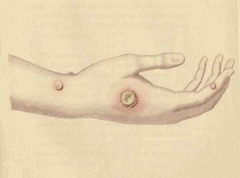
Fig. 1. Jenner’s drawing of cowpox lesion from which he created his vaccine.
Koch, Pasteur, and the germ theory of disease
In 1875, Robert Koch, a country physician with no formal scientific training, inoculated the ear of a rabbit with the blood of an animal that had died of anthrax. The rabbit died the next day. He isolated infected lymph nodes from the rabbit and was able to show that the bacteria contained within them could transfer disease to other animals. He developed and refined techniques necessary for the cultivation of bacteria, including the development of agar growth medium. He was appointed to the Institute of Hygiene in Berlin, where his ultimate goal was to identify the organism responsible for the “White Death”--tuberculosis.
Quite independently, Louis Pasteur began his studies of the “chicken cholera bacillus.” In a serendipitous discovery, Pasteur inadvertently left a flask of the bacillus on the bench over the summer and inoculated 8 chickens with this “old but viable” stock of chicken cholera bacillus. He found that not only did the chickens not die, but they did not even appear ill! Pasteur said that the virulent chicken cholera bacillus had become attenuated by sitting on the bench over the summer months. The similarity between these results and those of and Jenner using vaccinia virus was immediately apparent to him. In honor of Jenner, Pasteur called his treatment vaccination. Pasteur later worked on anthrax and rabies and developed the first viable vaccine for anthrax and rabies.
Although Koch and Pasteur were contemporaries, they were intensely competitive and actually bitter enemies--of course, the outbreak of the Franco-Prussian war (1870) did nothing to cement their relationship. In a trenchant example of how not to behave toward a colleague at a scientific meeting, Koch made his way to the podium following Pasteur’s lecture and said: “When I saw in the program that Monsieur Pasteur was to speak today...I attended the meeting eagerly, hoping to learn something new...I must confess that I have been disappointed, as there is nothing new in the speech which Monsieur Pasteur has just made...”
Although many consider Pasteur the “father of immunology” (?parent of immunology) it is due to both his and Koch’s efforts that firmly established the germ theory of disease. Prior to this time, although the practical benefits of variolation were apparent, there was no known biological basis for either the cause of diseases or the efficacy of vaccination.
The emerging distinction between cellular and humoral immunity
Metchnikoff was the first to recognize the contribution of phagocytosis to the generation of immunity. In Italy, while studying the origin of digestive organs in starfish larvae, he observed that certain cells unconnected with digestion surrounded and engulfed carmine dye particles and splinters that he had introduced into the bodies of the larvae. He called these cells phagocytes (from Greek words meaning “devouring cells”). Working first at the Bacteriological Institute in Odessa (1886-87), and later at the Pasteur Institute in Paris, Metchnikoff established that the phagocyte is the first line of defense against infection. He became a leading proponent of the “Cellularists” who believed that phagocytes, rather than antibodies, played the leading role in immunity.
Supporters of the alternative theory, the “Humoralists,” believed that a soluble substance in the body was mainly responsible for mediating immunity. Building upon the demonstration by Von Behring and Kitasato of the transfer of immunity against Diphtheria by a soluble “anti-toxin” in the blood, Paul Ehrlich predicted the existence of immune bodies (antibodies) and side-chains from which they arise (receptors). Ehrlich suggested that antigens interact with receptors borne by cells, resulting in the secretion of excess receptors (antibodies). Ehrlich surmised that erythrocytes would not have this capacity and speculated that this immune function might be a specialized characteristic or “haemopoietic tissue” (Fig. 2).
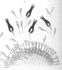
Fig. 2. Ehrlich’s drawing of a “haemopoietic” cell bearing “side chains” (receptors) and releasing “immune bodies” (antibodies).
Ehrlich was probably the first scientist to introduce the concept of immunological self/not-self discrimination, a mechanism “...which prevents the production within the organism of amboceptors (antibodies) directed against its own tissues. In this horror autoxicus, we are dealing with a well-adapted regulatory contrivance.”
Summary of the state of immunology at the end of the 19th century
By the turn of the century, several paradigms had been established that laid the groundwork for future studies in immunology. The first was based on the “germ theory” of disease (Koch and Pasteur) which held that disease was caused by bacteria. The second paradigm was that immunity to infection could be transferred by a soluble substance in the serum (Von Behring and Kitasato) elaborated by specialized cells of the immune system (Ehrlich) and that the regulation of this process (generation of antibodies) was important to minimize the possibility of developing an immune response against self (Ehrlich). Finally, the immune system responds to bacterial pathogens by the recruitment of “phagocytes,” which recognize, engulf, and destroy the microbes via “phagocytosis” (Metchnikoff). The first Nobel prize in Physiology or Medicine was awarded to von Behring “for his work on serum therapy, especially its application against diphtheria, by which he has opened a new road in the domain of medical science and thereby placed in the hands of the physician a victorious weapon against illness and deaths.” Metchnikoff and Ehrlich shared the Nobel prize in 1908 “in recognition of their work on immunity.”
The early dominance of humoral theories of immunity and later emergence of theories of cellular immunity
Between the years 1900 and 1942 the “Humoralists” played a dominant role in immunology. There were several reasons for this, not the least of which was the demonstration that transfer of immunity could be accomplished by soluble factors later shown to be antibodies (Von Behring, Roux) and complement (Bordet). Furthermore, much of the phenomenology of immunopathology (e.g., the Arthus reaction, anaphylaxis, serum sickness, hemolytic anemia) could be associated with the activity of specific circulating antibodies. Indeed, no other basis for immunological specificity was recognized. The case for antibodies as the fundamental unit of immunity was strengthened by the ascendancy of immunochemistry, a term coined by Arrhenius. The chemistry of antigen-antibody reactions was uncovered largely by the development of the quantitative precipitin reactions by Michael Heidelberger and Elvin Kabat (a former Professor at The College of Physicians & Surgeons). These studies paved the way for a more fundamental understanding of the immunoglobulin molecule, which culminated in the elucidation of antibody structure by Rodney Porter and Gerard Edelman in the late 1950s.
However, several experimental observations challenged the prevailing view that antibodies alone served to confer specific immunity. Delayed type hypersensitivity (e.g., tuberculin reactivity), first recognized by Koch in 1883, and allograft rejection (Medawar, 1944) appeared to be unrelated to the presence of circulating antibodies. The definitive proof that cells played a role in immunity came from the classic experiments of Landsteiner and Chase, in 1942. Cells from guinea pigs, which had been immunized with Mycobacterium tuberculosis or hapten, were transferred into naive guinea pigs. Later, when antigen or hapten was injected into these guinea pigs, they elicited an immune recall response that was not present in the naive controls. This did not happen when the serum fraction was transferred. Similar results were obtained using a contact dermatitis model. Thus, the dichotomy of immediate (antibody-mediated) and delayed-type (cell-mediated) hypersensitivity had become firmly established by the 1940s, although the
the identity of the cell that conferred the latter response was unknown. It was not until the pioneering experiments of Gowans that lymphocytes were recognized as being essential to immunity (Gowans et al., Initiation of immune responses by small lymphocytes. Nature 196:651-55, 1962). In the meantime, the genetic basis for the immune response, and its ontogeny, were gradually uncovered during the 1950s and 1960s.
A major paradigm shift: the clonal selection theory as an explanation for the diversity of the antibody repertoire
Prior to the 1950s, it was not known how antibody diversity was generated. Because the variability of antibodies was so great, early theories assumed that antibodies could not be preformed; rather, they would be synthesized on demand following exposure. It was therefore suggested that antigen instructs the cell about the specificity of the antibody. Indeed, in 1956, Burnet himself published a book maintaining the position that an antigen directs, rather than selects, the formation of specific antibody. In the late 1950s, three scientists (Jerne, Talmage, Burnet), working independently, developed what is widely referred to as the clonal selection theory. In 1955, Jerne published a paper (The natural-selection theory of antibody formation. Proc. Nat. Acad. Sci. 41: 849-857, 1955) that described a “selective” hypothesis, which held that every animal had a large set of natural globulins that had become diversified in some unknown fashion According to Jerne, the function of an antigen was to combine with those globulins with which it made a chance fit. The antigen would serve to transport the selected globulins to antibody-producing cells, which would then make many identical copies of the globulin presented to them. This was a seminal paper in the history of immunology, which presaged the key 1957 publications of Talmage (Allergy and immunology. Annu. Rev. Med. 8, 239-256, 1957) and Burnet (A modification of Jerne’s theory of antibody production using the concept of clonal selection. Aust. J. Sci. 20, 67-69, 1957).
In 1957, Talmage wrote:
“...it is tempting to consider that one of the multiplying units in the antibody response is the cell itself. According to this hypothesis, only those cells are selected for multiplication whose synthesized product has affinity for the antigen injected. This would have the disadvantage of requiring a different species of cell for each species of protein produced, but would not increase the total amount of configurational information required on the hereditary process.”
The evidence he cited to support his theory included the kinetics of the antibody response, the existence of “immunological memory” and the ability of myeloma tumors to secrete massive amounts of “one globulin randomly selected from the family of normal globulins.”
According to Burnet, the clonal selection theory states:
1. Animals contain numerous cells called lymphocytes.
2. Each lymphocyte is responsive to a particular antigen by virtue of specific surface receptor molecules.
3. Upon contacting its appropriate antigen, the lymphocyte is stimulated to proliferate (clonal expansion) and differentiate.
4. The expanded clone is responsible for the secondary response (more cells to respond) while the differentiated (“effector”) cells secrete antibody.
On the basis of many experiments in the ensuing years, the clonal selection theory was proven to be correct (Fig. 3). In 1960, along with Peter Medawar, Burnet was awarded the Nobel prize, “for discovery of acquired immunological tolerance” rather than the clonal selection theory. Jerne would later win the Nobel prize in 1984 “for theories concerning the specificity in development and control of the immune system.” Although Talmage received numerous awards, he did not receive the Nobel prize.
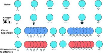
Fig. 3. The Clonal selection theory of lymphocytes. Schematic of clonal selection hypothesis illustrating the idea that each naïve lymphocyte has a different receptor specificity, each of which can bind a different antigenic determinant. When a pathogen is recognized by the cells, in this case by two different antigenic determinants, then the cells that bind to these determinants are selected to proliferate or undergo clonal expansion, and then differentiate into effector cells that either secrete antibody or mediate various effector mechanisms of cell-mediated immunity. From Abbas and Janeway Cell 100:129, 2000.
How lymphocytes function: the discovery of the Major Histocompatibility Complex and the emerging science of transplantation
The clonal selection theory represented a conceptual breakthrough in the history of immunology, but it did not explain how lymphocytes actually recognize antigen. These insights would eventually come principally from two sources: studies of the genetics of graft rejection in inbred strains of mice by Snell in the 1930s and studies of the agglutination of white blood cells by sera from transfused patients by Dausset in the 1950s. Snell was interested in tumor genetics and observed that tumor grafts were accepted between inbred mice, but not between mice of different strains. The same was true for normal tissues. Snell termed the underlying genes histocompatibility genes. In collaboration with Peter Gorer, Snell established that the major locus was identical to a locus encoding antigen II, renamed locus histocompatibility 2, or H-2. Analogously, Dausset observed that patients, who had received many blood transfusions, produced antibodies that could agglutinate white blood cells from donors, but not the patient’s own cells. Subsequent family studies indicated a genetically determined system, termed HLA, was found to the ortholog of H-2 in the mouse. Research in mice and humans became mutually complementary and the Nobel Prize was awarded to Dausset in 1980, together with Snell and Benaceraff.
The discovery of the MHC had widespread implications, paricularly in the field of organ transplantation. The impetus for the study of transplantation biology came from the war, which led to a marked increase in the number of burn victims. In many of these individuals, a skin autograft was not feasible. The application of skin grafts from other individuals (allografts) was known for its high failure rate, due to rejection. The British Medical Council assigned a young Oxford-trained zoologist named Peter Medawar to investigate the problem of graft rejection. In 1943, Medawar and Gibson published “The Fate of Skin Homografts in Man” based on a single burn victim who received multiple “pinch grafts” of skin. The authors concluded that autografts succeed, while allografts fail after an initial take, and that the destruction of the foreign epidermis is brought about by a mechanism of active immunization. Medawar returned to Oxford University to study the homograft rejection in laboratory animals and proved that this was an immunologic phenomenon.
Medawar concluded that the mechanism by which foreign skin is eliminated belongs to the category of “actively acquired immune reactions.” His early insight into the mechanism of transplant rejection is reflected by statements such as “The accelerated regression of second-set homografts argues for the existence of a systemic immune state.”
The concept of immunological tolerance and the “self-nonself” model of immune development
The discovery of neonatal transplant tolerance has been credited to Ray Owen, a geneticist at the University of Wisconsin who studied the inheritance of red blood cell antigens in cattle. He reported in 1945 that dizygotic twins had mixtures of their own cells and their twin partner cells. Owen recognized that the common intrauterine circulation of cattle leads to an exchange of hematopoietic stem cells during embryonic life and the establishment of a chimeric state of red cells. These calves did not develop antibodies to their twin partners. A few years later, Burnet and Fenner acknowledged in their influential book “The Production of Antibodies” the importance of Owen’s findings, which led to the “self-nonself” hypothesis for immune development. They postulated that, during embryonic development, “a process of self-recognition takes place” and “no antibody response should develop against the foreign cell antigen when the animal takes on independent existence.” Owen’s red cell chimeric model in dizygotic cattle and Burnet’s own studies of foreign embryonic cells in the chick embryo led Burnet to hypothesize that the existence of “tolerance acquired by fetal exposure to ‘nonself’ constituents.”
Medawar predicted that an exchange of skin grafts between dizygotic calves would verify Burnet’s hypothesis. Together with his post-doctoral fellow Rupert Billingham, he performed a series of grafting experiments that provided direct support for the concept of neonatally-acquired transplantation tolerance. At the same time, Milan Hasek in Prague demonstrated that parabiosis of different strain chick embryos induced a immune hyporesponsive state to each other’s red cells.
The discovery of MHC restriction as the genetic basis for “self-nonself” recognition
In 1974, Peter Doherty and Rolf Zinkernagel sought to learn the role of T lymphocytes (T-cells) in the immune response to viral meningitis. They theorized that it was the strength of the immune response that caused the fatal destruction of brain cells infected with this virus. To test this theory, they mixed virus-infected mouse cells with T lymphocytes from other infected mice. The T lymphocytes did destroy the virus-infected cells, but only if the infected cells and the lymphocytes came from a genetically identical strain of mice. T lymphocytes would ignore virus-infected cells that had been taken from another strain of mice (Zinkernagel RM, Doherty PC Restriction of in vitro T cell-mediated cytotoxicity in lymphocytic choriomeningitis within a syngeneic or semiallogeneic system. Nature 248:701-2, 1974); (Fig. 3).
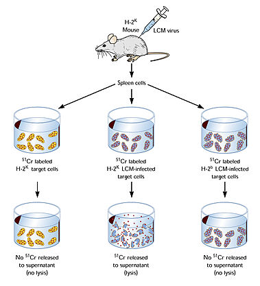
Fig. 3. Zinkernagel-Doherty experiment demonstrating that MHC restriction is required for activation of a cytotoxic T-cell response.
The implications of the Zinkernagel-Doherty experiment were profound. First, it established the principle of MHC restriction: T cells recognize antigen only in the context of MHC molecules. It would take another 13 years to prove that the antigen in question was a peptide actually bound to the MHC molecule. Second, the experiment established that cells must recognize two separate signals on an infected cell before they can destroy it. One signal is a fragment of the invading virus that the cell displays on its surface; the other is a self-identifying tag from the cell’s major histocompatibility complex (MHC) antigens. Thus, the experiment pointed to the identity of the molecular structure that constituted immunological “self”--it is the MHC molecule; a virus-infected cell bearing MHC molecules constitutes “altered-self.” Finally, the fact that MHC is highly polymorphic (i.e., multiple alleles expressed in different individuals) implies that any one allele may respond differently to a given stimulus; thus, the specific identity of the MHC molecule itself determines the strength of the immune response.
The generation of immunological diversity
Because the immune system has the capacity to respond to a multitude of environmental insults, it must have an efficient way of insuring the diversity of its responses; this is sometimes referred to the T- or B-cell “repertoire.” The mechanism by which this was accomplished remained elusive until 1978, when Susumu Tonegawa provided direct evidence for somatic rearrangement of immunoglobulin genes. This represented a radical departure from one of the fundamental dogmas of molecular genetics, which held that the genetic makeup of an organism remained unchanged throughout ontogeny (unless altered by pathological states, such as cancer). Indeed, the immunoglobulin genes and the genes that make up the T-cell antigen receptor are the only genes that have been shown to undergo somatic rearrangements. The various combinations of genetic elements within these loci accounts for much of the diversity of the T- and B-cell repertoire, although other mechanisms, such as somatic hypermutation, would later be discovered to generate further diversity.
The complementary roles played by cellular and molecular immunology
Since 1974, much progress has been made in uncovering precisely how antigens are recognized by the immune system. These insights have come from two complementary approaches: a molecular one, involving the cloning of the genes for the T-cell antigen receptor (1984-87) and solving the crystal structure of peptide bound to the MHC molecule (1987), and a cellular one, delineating the cellular mechanisms of antigen presentation, leukocyte trafficking, and signal transduction. In 1978, Ralph Steinman identified the dendritic cell, a phagocytic cell, as the principal antigen-presenting cell of the immune system. This constituted a major revision to the role of the phagocyte assigned by Metchnikoff in the 19th century! Other advances included the identification of adhesion molecules (Butcher, 1979) and chemokines (Leonard, Yoshimura, and Baggiolini,1989) together which provide the cellular basis for leukocyte trafficking.
Since 1986, a major effort has been directed towards identifying markers of individual T-cell subsets (“phenotyping”) and characterizing their function. In 1986, Tim Mossmann and Bob Coffman discovered a major dichotomy in T helper subsets: TH1 cells, which are responsible for the production of interferon-g and the activation of macrophages (as well as the principal lymphocyte effector of delayed-type hypersensitivity), and TH2 cells, which are required for the production of certain types of immunoglobulins and are implicated in the pathogenesis of allergic diseases and immediate hypersensitivity. These specific T cell subsets elaborate a distinct array of soluble substances that influence the behavior of other cells (“cytokines”). The search for cytokines began in 1957 with the discovery of interferon (Issacs and Lindemann), but the characterization of the properties of various cytokines is an ongoing enterprise.
Progress in cellular immunology leads to insights into T-cell effector functions: “two signal models” for lymphocyte activation
The origins of cellular immunology date back to Metchnikoff, who discovered the important role that phagocytes play in immunity. Although he mistakenly identified the phagocyte, itself, as the cell type responsible for antibody production, it would take another 58 years before any further understanding of the cellular basis of immunity would exist. In 1942, Landsteiner and Chase demonstrated that cells, rather than antibodies, were recognized to contribute to specific immune phenomena. As there was no technology available to separate different types of circulating leukocytes, the nature of the immune-conferring cell remained elusive. It was not until the experiments of Gowans in 1959 that lymphocytes were shown to confer specific immunity. In many ways, Gowans’ discovery of the central role of lymphocytes in immunity was analogous to the molecular genetic experiments of Avery, McLeod, and Lederberg, who demonstrated that DNA was the substance that conferred heredity.
The focus on the lymphocyte led to the discovery of the involvement of the thymus in cellular immunity and the distinction between T-cells, which provided “help” or cytotoxicity, and B-cells, which produced antibody (Jacques Miller and Graham Mitchell, 1961-1968). These workers also discovered the importance of T- and B-cell collaboration in the immune response. In 1968, Bretscher and Cohn proposed the first two-signal model of lymphocyte activation. Although the original model proved to be incorrect, the model has been refined over the years to take into account new experimental findings.
The two signal hypothesis basically states that, in addition to the delivery of antigen/MHC-mediated signals to the lymphocyte, there must be another signal provided by the MHC-bearing antigen-presenting cell (Fig. 4 ). The nature of this “second stimulus” was unknown until the
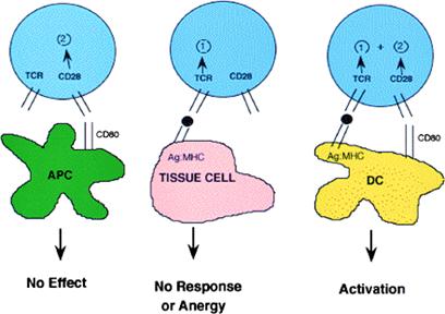
Fig. 4. The Two-signal theory of T-cell activation. When a T-cell encounters an antigen presenting cell (APC) that expresses costimulatory molecules but no foreign antigens, there is no apparent signal (left panel); when a T cell encounters a tissue cell or an APC that expresses antigens recognized by the TCR in the absence of costimulatory signals, the result is either no response or inactivation of the cell (anergy). However, when a T cell encounters an activated dendritic cell or other APC expressing both antigen and costimulatory molecules, then the T cell is activated to proliferate and undergo differentiation to an effector cell. Thus, T cells only become activated by their ligands when they are presented by a cell that expresses costimulatory molecules. From Abbas and Janeway Cell 100:129, 2000.
the interaction of CD28 on the T-cell and B7 (CD80/CD86) on the antigen-presenting cell was necessary for optimal production of antibody. Later studies (1992) identified CD40 ligand (CD154) on activated T-cell as a critically important molecule that provides co-stimulation to B-cells; in humans. In the following year, it was demonstrated that mutations in the gene for CD40 ligand was responsible for a human disease, X-linked hyper-IgM immunodeficiency.
However, even before the precise identity of co-stimulatory molecules was known, a theoretical framework for T-cell function was proposed by Burnet (1959) and later refined over the ensuing years. These models for T-cell function incorporated the “two signal” concept of lymphocyte activation, originally proposed by Bretscher and Cohn, into a model of immune recognition based on “self vs non-self.” (Table I).
Table I. Evolution of “Two Signal” Model of Lymphocyte Activation

*Pattern recognition receptor
How valid are these models? There is no doubt that the activation of T-cells requires multiple signals from antigen-presenting cells. Where the models diverge, and where the most controversy exists, is the very basis of immune recognition. What constitutes “non-self”? Because the immune response in any individual depends upon the selection of a small subset of lymphocytes out of a total of 105 to 106 possible clones, the very existence of these clones implies that they were positively selected by the immune system. But this occurs through an ongoing low-level presentation of self antigens, thus constituting a paradox: recognition of “non-self” depends on prior recognition of “self.” How can this be resolved?
The role of innate immunity in the acquired immune response
The components of acquired immunity (i.e., lymphocytes, immunoglobulins, MHC molecules, antigen receptors) are absent in primitive species. Genes for these proteins appeared abruptly in evolution with the advent of lower vertebrates. Recognizing the presence of components of primitive or “innate” immunity in higher organisms, Janeway proposed in 1989 an alternative to “self/nonself” recognition by the immune system. He suggested that the immune system recognizes “infectious non-self.” Early in an immune response, when components of innate immunity are called into play, lymphocytes and/or antigen presenting cells are stimulated to respond in a stereotypic fashion that serves to initiate the acquired immune response. Janeway predicted the existence of “pattern recognition receptors” that recognize products of microbial pathogens that differed from “self” in that they bore repetitive structures (e.g., components of bacterial cell walls) that were not present in the host. Although there was as yet no formal proof for this highly original concept, in 1997, Janeway and Medzhitov identified a human homolog of a transmembrane protein in Drosophila, Toll, that conferred responsiveness to lipopolysaccharide, a component derived from the cell wall of gram-negative bacteria. Furthermore, ectopic expression of this receptor resulted in the secretion of a pro-inflammatory cytokine and resulted in the up-regulation of co-stimulatory molecules known to be important in triggering an acquired immune response in T-cells.
The discovery of the components of the innate immune system, and their likely role in triggering acquired immunity, represents a paradigm shift in immunology perhaps as profound as the clonal selection theory. Since Janeway proposed this in 1989, Matzinger suggested that, rather than sensing pathogen-derived signals directly, the immune system senses “danger” that results as a consequence of infection. This danger can be in the form of injured or dying cells; indeed, several proteins released from necrotic cells, including heat-shock proteins, have been demonstrated to stimulate antigen-presenting cells, such as macrophages. Interestingly, the same receptors on macrophages that respond to bacterial products (Toll-like receptors) are also required for the response to heat shock proteins!
The future of immunology?
"Predictions are hard to make, especially ones about the future" -Peter Medawar
It is pure speculation (but fun) to guage the direction that immunology is headed in the next 20-30 years. Further advances in the cell biology of the immune system will no doubt occur, which will lead to novel vaccines for infectious and non-infectious diseases, such as cancer and diseases of aging. New receptor- or cytokine-modifying therapeutics will be developed, based on insights obtained from experimental immunology. The application of the human genome project to diseased populations will identify new drug targets, and high-throughput screens and combinatorial chemistry will accelerate the pace of drug discovery. Gene and protein microarray techniques and proteomics will reveal new components of immunity that will expand our knowledge of how the immune system works. Perhaps more importantly, we will begin to appreciate the fundamental role that the immune system plays in nearly all human diseases, and exploit this knowledge to alter the natural history of these diseases.


|
|
|
|
التوتر والسرطان.. علماء يحذرون من "صلة خطيرة"
|
|
|
|
|
|
|
مرآة السيارة: مدى دقة عكسها للصورة الصحيحة
|
|
|
|
|
|
|
نحو شراكة وطنية متكاملة.. الأمين العام للعتبة الحسينية يبحث مع وكيل وزارة الخارجية آفاق التعاون المؤسسي
|
|
|