


النبات

مواضيع عامة في علم النبات
الجذور - السيقان - الأوراق
النباتات الوعائية واللاوعائية
البذور (مغطاة البذور - عاريات البذور)
الطحالب
النباتات الطبية
الحيوان
مواضيع عامة في علم الحيوان
علم التشريح
التنوع الإحيائي
البايلوجيا الخلوية
الأحياء المجهرية
البكتيريا
الفطريات
الطفيليات
الفايروسات
علم الأمراض
الاورام
الامراض الوراثية
الامراض المناعية
الامراض المدارية
اضطرابات الدورة الدموية
مواضيع عامة في علم الامراض
الحشرات
التقانة الإحيائية
مواضيع عامة في التقانة الإحيائية
التقنية الحيوية المكروبية
التقنية الحيوية والميكروبات
الفعاليات الحيوية
وراثة الاحياء المجهرية
تصنيف الاحياء المجهرية
الاحياء المجهرية في الطبيعة
أيض الاجهاد
التقنية الحيوية والبيئة
التقنية الحيوية والطب
التقنية الحيوية والزراعة
التقنية الحيوية والصناعة
التقنية الحيوية والطاقة
البحار والطحالب الصغيرة
عزل البروتين
هندسة الجينات
التقنية الحياتية النانوية
مفاهيم التقنية الحيوية النانوية
التراكيب النانوية والمجاهر المستخدمة في رؤيتها
تصنيع وتخليق المواد النانوية
تطبيقات التقنية النانوية والحيوية النانوية
الرقائق والمتحسسات الحيوية
المصفوفات المجهرية وحاسوب الدنا
اللقاحات
البيئة والتلوث
علم الأجنة
اعضاء التكاثر وتشكل الاعراس
الاخصاب
التشطر
العصيبة وتشكل الجسيدات
تشكل اللواحق الجنينية
تكون المعيدة وظهور الطبقات الجنينية
مقدمة لعلم الاجنة
الأحياء الجزيئي
مواضيع عامة في الاحياء الجزيئي
علم وظائف الأعضاء
الغدد
مواضيع عامة في الغدد
الغدد الصم و هرموناتها
الجسم تحت السريري
الغدة النخامية
الغدة الكظرية
الغدة التناسلية
الغدة الدرقية والجار الدرقية
الغدة البنكرياسية
الغدة الصنوبرية
مواضيع عامة في علم وظائف الاعضاء
الخلية الحيوانية
الجهاز العصبي
أعضاء الحس
الجهاز العضلي
السوائل الجسمية
الجهاز الدوري والليمف
الجهاز التنفسي
الجهاز الهضمي
الجهاز البولي
المضادات الميكروبية
مواضيع عامة في المضادات الميكروبية
مضادات البكتيريا
مضادات الفطريات
مضادات الطفيليات
مضادات الفايروسات
علم الخلية
الوراثة
الأحياء العامة
المناعة
التحليلات المرضية
الكيمياء الحيوية
مواضيع متنوعة أخرى
الانزيمات

Four-Helix Bundle Motif
 المؤلف:
J. Trikha, E. C. Theil, and N. M. Allewell
المؤلف:
J. Trikha, E. C. Theil, and N. M. Allewell
 المصدر:
J. Mol. Biol. 248, 949–967
المصدر:
J. Mol. Biol. 248, 949–967
 الجزء والصفحة:
 11-5-2016
 2751
2751


+
-
20
Four-Helix Bundle Motif
The four-helix bundle is a common protein motif that is observed in protein structures, in which four a-helices pack together lengthwise, in an antiparallel manner (Fig. 1). The four a-helices that form the bundle can derive from four independent polypeptide chains or from a single polypeptide chain. This structural motif has been observed both as an isolated three-dimensional fold and as a domain within much larger and more complex protein structures. The helices of four-helix bundles are longer than average (~20 residues, as opposed to the usual 10) and are amphipathic, with hydrophobic residues buried in the core. The antiparallel packing of the helices in the bundle may be favored by interaction between the helix dipoles.
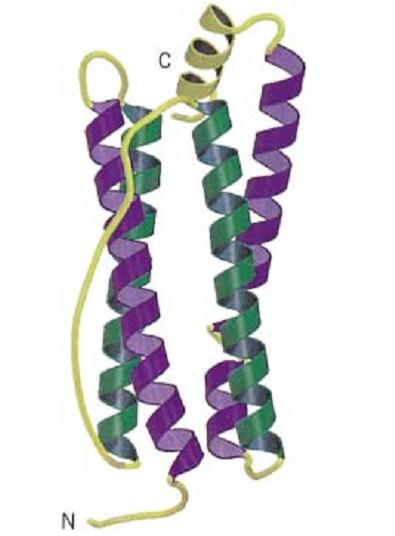
Figure 1. Schematic representation of the backbone of a four-helix bundle protein, taken from the structure of bullfrog ferritin (1). This structure has the up–down–down–up topology; the two “up” helices are shown as purple coils and the two “down” helices are shown as green coils. Note the long connecting loop between the second and third helix. Connecting loops and an additional C-terminal helix are shown in yellow. The N- and C-termini are labeled. This figure was generated using Molscript (2) and Raster3D (3, 4).
The four-helix bundle is one of the simplest protein folds and is used by a wide range of proteins with diverse functions. Proteins with a four-helix bundle structure include growth hormones, cytokines, and ferritins. The four helices may splay apart to generate a binding pocket for cofactors and metal ions, or they may coil around each other to form a supercoil. In the latter case, the twist of the helices in the coiled coils alters the usual helical structure so that there are 3.5 residues per turn, rather than 3.6. This means that every seventh residue in the helix is identically placed with respect to the axis of the helix. The seven positions of this heptad repeat are denoted a to g, and residues a and d point into the core of the bundle and are usually hydrophobic.
Several classes of four-helix bundle motif have been defined. These include (a) the simple up–down–up–down topology, where consecutive helices have short connections and alternating directions, (b) the up-down-down-up topology, where there is a long loop or crossover connection between helices 2 and 3, and (c) the up–up–down–down topology that is typical of the cytokine four-helix bundles, with two long crossover connections between helices 1 and 2 and helices 3 and 4. In most cases, the helices are aligned so that each pairwise interaction is antiparallel. There are also sub-classes within each of these classifications. The simplicity of the four-helix bundle motif has made it a common target for protein design.
References
1. J. Trikha, E. C. Theil, and N. M. Allewell (1995) J. Mol. Biol. 248, 949–967.
2. P. J. Kraulis (1991) J. Appl. Crystallogr. 24, 946–950.
3. E. A. Merritt and M. E. P. Murphy (1994) Acta Crystallogr. D50, 869–873.
4. D. J. Bacon and W. F. Anderson (1988) J. Mol. Graphics 6, 219–222.
 الاكثر قراءة في مواضيع عامة في الاحياء الجزيئي
 اخر الاخبار
اخبار العتبة العباسية المقدسة

الآخبار الصحية


مواضيع ذات صلة
 قسم الشؤون الفكرية يصدر كتاباً يوثق تاريخ السدانة في العتبة العباسية المقدسة
 "المهمة".. إصدار قصصي يوثّق القصص الفائزة في مسابقة فتوى الدفاع المقدسة للقصة القصيرة
 (نوافذ).. إصدار أدبي يوثق القصص الفائزة في مسابقة الإمام العسكري (عليه السلام)


