

 النبات
 الحيوان
 الأحياء المجهرية
 علم الأمراض
 التقانة الإحيائية
 التقنية الحيوية المكروبية
 التقنية الحياتية النانوية
 علم الأجنة
 الأحياء الجزيئي
 علم وظائف الأعضاء
 الغدد
الغدد
 المضادات الحيوية
|
Read More
Date: 4-1-2017
Date: 3-1-2017
Date: 23-1-2017
|
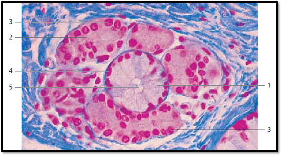
Extraepithelial Glands-Mixed (Seromucous) Glands
This figure displays a mucous terminal portion (tubulus) 1 . It is flanked by several serous acini 2 . The nuclei of serous acinar cells are round 3 while the nuclei of mucous tubule are more flattened 4 . The cytoplasm of serous gland cells is stained red, that of mucous gland cells is light. Note the wide lumen 5 of the mucous tubule.
1 Mucous tubule
2 Serous acini
3 Nuclei of serous gland cells
4 Nuclei of mucous gland cells
5 Lumen of a mucous tubule
Mixed glands from the mucous membrane of the uvula.
Stain: azan; magnification: × 400

References
Kuehnel, W.(2003). Color Atlas of Cytology, Histology, and Microscopic Anatomy. 4th edition . Institute of Anatomy Universitätzu Luebeck Luebeck, Germany . Thieme Stuttgar t · New York .


|
|
|
|
لصحة القلب والأمعاء.. 8 أطعمة لا غنى عنها
|
|
|
|
|
|
|
حل سحري لخلايا البيروفسكايت الشمسية.. يرفع كفاءتها إلى 26%
|
|
|
|
|
|
|
في مدينة الهرمل اللبنانية.. وفد العتبة الحسينية المقدسة يستمر بإغاثة العوائل السورية المنكوبة
|
|
|