آخر المواضيع المضافة

 النبات
الحيوان
الأحياء المجهرية
علم الأمراض
التقانة الإحيائية
التقنية الحيوية المكروبية
التقنية الحياتية النانوية
علم الأجنة
الأحياء الجزيئي
علم وظائف الأعضاء
الغدد
المضادات الحيوية
النبات
الحيوان
الأحياء المجهرية
علم الأمراض
التقانة الإحيائية
التقنية الحيوية المكروبية
التقنية الحياتية النانوية
علم الأجنة
الأحياء الجزيئي
علم وظائف الأعضاء
الغدد
المضادات الحيوية| Optic Nerve |


|
|
Read More
Date: 10-1-2017
Date: 26-7-2016
Date: 11-1-2017
|
Optic Nerve
Section of the optic nerve behind the lamina cribrosa sclerae.
1 Ciliary nerve
2 Sclera
3 Pigment epithelium of the lamina suprachoroidea
4 Fiber bundles of the optic nerve
5 Central retinal artery
6 Sept a of the piamater
7 Central retinal vein
8 Internal sheath of the optic nerve (piamater)
9 External sheath of the optic nerve (dura mater)
10 Subdural space
11 Arachnoidea
12 Short posterior ciliary artery
Stain: hematoxylin-eosin; magnification: × 20
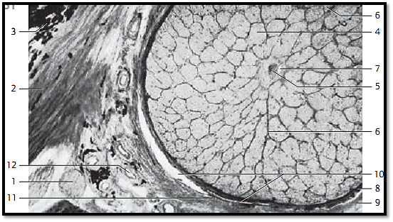
Optic Nerve
Cross-section of the optic nerve behind the lamina cribrosa sclerae. The axons of the ganglia cells are combine d in bundles 1 . They are envelope d by a thin septum of the piamater 2 . The optic nerve contains a piamater sheath, an arachnoidea sheath and a dura mater sheath. The piamater sheath attaches directly to the optic nerve. Septa originate with the piamater and guide blood vessels to the myelinate nerve f ibers. The nerve fiber bundles contain astrocytes and oligodendrocytes. Arteries 3 and central retinal veins 4 are visible in the center of the section. The vessels are sheathed by the loose connective tissue of the piamater.
1 Nerve fiber bundles
2 Piamater septa
3 Central retinal arter y
4 Central retinal vein
Stain: hematoxylin-eosin; magnification: × 40
References
Kuehnel, W.(2003). Color Atlas of Cytology, Histology, and Microscopic Anatomy. 4th edition . Institute of Anatomy Universitätzu Luebeck Luebeck, Germany . Thieme Stuttgart · New York .


|
|
|
|
تحذير من "عادة" خلال تنظيف اللسان.. خطيرة على القلب
|
|
|
|
|
|
|
دراسة علمية تحذر من علاقات حب "اصطناعية" ؟!
|
|
|
|
|
|
|
العتبة العباسية المقدسة تحذّر من خطورة الحرب الثقافية والأخلاقية التي تستهدف المجتمع الإسلاميّ
|
|
|