

 النبات
 الحيوان
 الأحياء المجهرية
 علم الأمراض
 التقانة الإحيائية
 التقنية الحيوية المكروبية
 التقنية الحياتية النانوية
 علم الأجنة
 الأحياء الجزيئي
 علم وظائف الأعضاء
 الغدد
الغدد
 المضادات الحيوية
|
Read More
Date: 10-7-2021
Date: 12-7-2021
Date: 30-7-2021
|
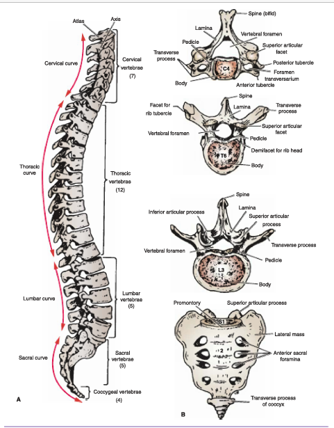
The sacrum (see Fig. 1) consists of five rudimentary vertebrae fused together to form a wedge-shaped bone, which is concave anteriorly. The upper border, or base of the bone articulates with the fifth lumbar vertebra.

FIG 1 / Posterior view of the back showing the vertebral column and related surface markings.
The narrow Inferior border, or apex, articulates with the coccyx. Laterally, the sacrum articulates with the two iliac bones to form the sacroiliac joints (see Fig. 2).

FIG 2 / A. Lateral view of the vertebral column. B. General features of different kinds of vertebrae.
The anterior and upper margin of the first sacral vertebra bulges forward as the posterior margin of the pelvic Inlet and ls termed the sacral promontory. The sacral promontory in the female ls of considerable obstetric importance and is used when measuring the size of the pelvis.
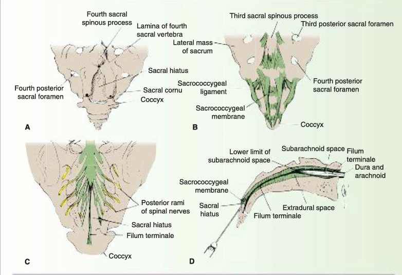
The vertebral canal continues into the sacrum where it forms the sacral canal. The sacral canal contains the cauda equina (composed mainly of the anterior and posterior roots of the sacral and coccygeal spinal nerves) and fibrofatty material. It also contains the lower part of the subarachnoid space down as far as the second sacral vertebra. The laminae of the fifth sacral vertebra. and sometimes those of the fourth, fan to meet in the midline, forming the sacral hiatus (see Fig. 3). At the sacral hiatus, the posterior inferior end of the sacral canal Is devoid of a bony cover, making the canal easily accessible .

FIG 3 / A. The sacral hiatus. Black dots indicate the position of important bony landmarks. B. Posterior surface of the lower end of the sacrum and the coccyx showing the sacrococcygeal membrane covering the sacral hiatus. C. The dural sheath (thecal sac) around the lower end of the spinal cord and spinal nerves in the sacral canal; the laminae have been removed. D. Longitudinal section through the sacrum showing the anatomy of caudal anesthesia.
The anterior and posterior surfaces of the sacrum each have four foramina (anterior and posterior sacral foramina) for the passage of the anterior and posterior rami of the upper four sacral nerves.
The fifth lumbar vertebra may be incorporated into the sacrum, in a condition termed sacralization on of the L5 vertebra. This is usually incomplete and may be limited to one side. Conversely, the first sacral vertebra may remain partially or completely separate from the sacrum and resemble a sixth lumbar vertebra (lumbarization of the S1 vertebra). A large extent of the posterior wall of the sacral canal may be absent because the laminae and spines fall to develop


|
|
|
|
بـ3 خطوات بسيطة.. كيف تحقق الجسم المثالي؟
|
|
|
|
|
|
|
دماغك يكشف أسرارك..علماء يتنبأون بمفاجآتك قبل أن تشعر بها!
|
|
|
|
|
|
|
لمجمع العلمي يقيم دورات ومحافل لتعزيز الثقافة القرآنية ونشر تعاليمها
|
|
|